Figures & data
Table 1 Clinical and pathological features of 127 study patients
Figure 1 Different expression levels of TRPV1 in normal kidney vs RCC.
Notes: (A) In quantitative RT-PCR, TRPV1 mRNA level was decreased 3.2-fold in the RCC vs kidney. (B) Western blot analysis reveals lower TRPV1 expression in RCC than in kidney samples. (C) Densitometric analysis of the TRPV1/β-actin protein bands in B. *A statistically significant difference of TRPV1 mRNA or protein levels in RCC vs normal peritumoral kidney tissues.
Abbreviations: OD, optical density; RCC, renal cell carcinoma; RT-PCR, reverse transcription polymerase chain reaction; TRPV1, transient receptor potential vanilloid type-1.
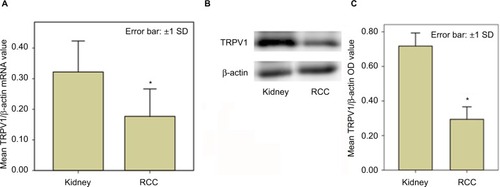
Figure 2 Quantitative RT-PCR compared TRPV1 mRNA expression among different histopathological subtype and Fuhrman grades of RCC.
Notes: (A) Significant differences were detected among chrRCC, gRCC and pRCC vs normal peritumoral kidney tissues of the same patients, but none were found in ccRCC vs kidney tissues. (B) Significant differences were also detected among G2RCC and G3RCC vs normal peritumoral kidney tissues in the same patients, but none were found in G1RCC. *A statistically significant difference (p<0.05).
Abbreviations: ccRCC, clear cell RCC; chrRCC, chromophobe RCC; gRCC, granular RCC; G1RCC, grade 1 RCC; G2RCC, grade 2 RCC; G3RCC, grade 3 RCC; pRCC, papillary RCC; RCC, renal cell carcinoma; RT-PCR, reverse transcription polymerase chain reaction; TRPV1, transient receptor potential vanilloid type-1.
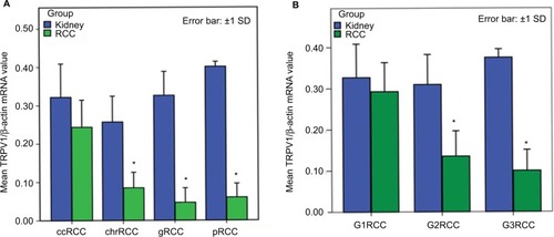
Figure 3 Western blot analysis compared TRPV1 protein expression among different histopathological subtype and Fuhrman grades of RCC.
Notes: (A) Western blot analysis showing relatively lower TRPV1 expression in RCCs than in normal peritumoral kidney tissues. Different expression levels were also detected among RCC histopathological subtypes. (B) OD ratios of kidney and different histopathological subtypes of RCC. (C) Relatively lower TRPV1 expression was detected in RCC samples with higher Fuhrman grades. (D) OD ratios of kidney and different Fuhrman grades of RCC. *Statistically significant difference (p<0.05).
Abbreviations: ccRCC, clear cell RCC; chrRCC, chromophobe RCC; gRCC, granular RCC; G1RCC, grade 1 RCC; G2RCC, grade 2 RCC; G3RCC, grade 3 RCC; OD, optical density; pRCC, papillary RCC; RCC, renal cell carcinoma; TRPV1, transient receptor potential vanilloid type-1.
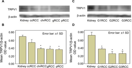
Figure 4 TRPV1 IHC staining in normal kidney tissue and RCC histopathological subtypes.
Notes: (A, B) Staining was moderate to strong in the proximal tubule cytoplasm and weak to moderate in CDs and DTs, but glomeruli was negative for TRPV1. (C) Diffusely moderate immunoreactivity in clear cell RCC. (D) Focal weak to moderate immunoreactivity in granular RCCs. (E, F) Focal weak to poor staining in chromophobe RCCs and papillary RCCs could be detected. Hematoxylin counterstain, reduced from ×200.
Abbreviations: CDs, collecting ducts; DTs, distal tubules; IHC, immunohistochemical; RCC, renal cell carcinoma; TRPV1, transient receptor potential vanilloid type-1.
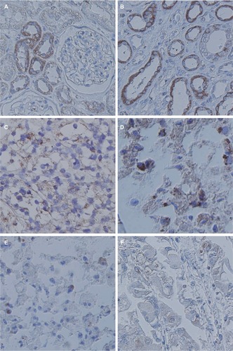
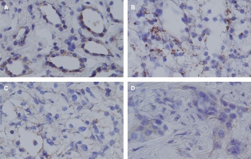
Figure 5 TRPV1 IHC staining in normal kidney and different ccRCC Fuhrman grades.
Notes: (A) TRPV1 strongly expressed in the renal tubules. (B) G1RCC showed focal/moderate to strong immunoreactivity. (C) G2RCC showed focal/weak immunoreactivity. (D) G3RCC showed weak or no positive staining. Reduced from ×200.
Abbreviations: ccRCC, clear cell RCC; G1RCC, grade 1 RCC; G2RCC, grade 2 RCC; G3RCC, grade 3 RCC; IHC, immunohistochemical; RCC, renal cell carcinoma; TRPV1, transient receptor potential vanilloid type-1.