Figures & data
Figure 1 PYCR1 expression was upregulated and predicted a poor prognosis in human MM.
Notes: The expression and prognosis role of PYCR1 were assessed by using the GEPIA online database. (A) PYCR1 expression was significantly increased in SKCM tumor tissues (n=461, red box) in comparison to normal skin tissue (n=558, gray box). (B) The survival curve was plotted in SKCM patients who were divided into PYCR1 high-expression group (n=229) and PYCR1 low-expression group (n=229). As seen from the curve, SKCM patients with low PYCR1 level showed better OS than those with high PYCR1 level. *P<0.05.
Abbreviations: MM, malignant melanoma; GEPIA, Gene Expression Profiling Interactive Analysis; SKCM, skin cutaneous melanoma; OS, overall survival; TPM, transcripts per million; PYCR1, pyrroline-5-carboxylate reductase 1.
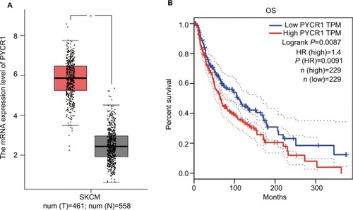
Figure 2 Knockdown of PYCR1 inhibited proliferation and migration in human MM cells.
Notes: PYCR1 expression was analyzed by using (A) qPCR and (B and C) Western blotting in three human melanoma cell lines, A375, M14 and UACC257, and in one human normal melanocyte cell line, HEM. PYCR1 level was upregulated in A375, M14 and UACC257 cell lines compared with HEM. (D) PYCR1 expression was significantly suppressed by PYCR1 siRNA in A375 and M14 cells. (E) CCK8 assay was performed to assess the proliferation of siPYCR1 and NC cells. After 72 hours of transfection, the OD value of siPYCR1 cells decreased significantly. (F and G) Cell migration was determined by transwell assay. The number of migrating cells significantly declined in the siPYCR1 group compared to the NC group. *P<0.05 and **P<0.01.
Abbreviations: MM, malignant melanoma; qPCR, quantitative PCR; CCK8, Cell Counting Kit-8; PYCR1, pyrroline-5-carboxylate reductase 1; siPYCR1, PYCR1-specific siRNA; HEM, human epidermal melanocyte cell line; NC, negative control.
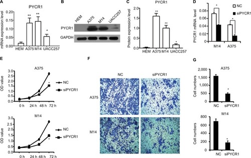
Figure 3 Knockdown of PYCR1 induced apoptosis in human MM cells.
Notes: (A) Flow cytometric analysis was performed to estimate cellular apoptosis. (B) The percentage of apoptosis in A375 cells transfected with siPYCR1 significantly increased to 18.71±1.01 in comparison to that of the NC group, 1.71±1.02. (C) The percentage of apoptosis in M14 cells transfected with siPYCR1 significantly increased to 23.79±2.52 in comparison to that of the NC group, 12.61±0.11. *P<0.05.
Abbreviations: MM, malignant melanoma; FITC, fluorescein isothiocyanate; PI, propidium iodide; PYCR1, pyrroline-5-carboxylate reductase; siPYCR1, PYCR1-specific siRNA; NC, negative control.
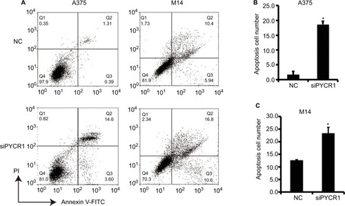
Figure 4 PYCR1 played a role in tumor progression of human MM through the stimulation of the AKT pathway.
Notes: (A) The expression of AKT signaling pathway-related genes was analyzed by Western blot. (B and C) Statistical analysis of Western blot results was performed using ImageJ software. siPYCR1 inhibited AKT phosphorylation and P70 level, and promoted the expression of the apoptosis factors, Caspase3-p17 and Bax in A375 and M14 cell lines. (D and E) Rescue experiments were performed by activating the AKT pathway with IGF-1 in PYCR1 interference cells (siPYCR1+ IGF-1). CCK8 was used to detect cell proliferation in three cell groups. (F) The expression of AKT signaling pathway-related genes was analyzed by Western blot. (G) Statistical analysis of Western blot results was performed using ImageJ software. *P<0.05.
Abbreviations: MM, malignant melanoma; CCK8, Cell Counting Kit-8; PYCR1, pyrroline-5-carboxylate reductase; siPYCR1, PYCR1-specific siRNA; NC, negative control.
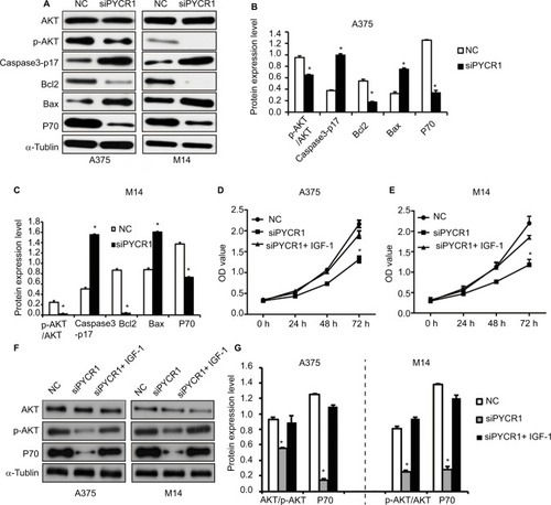
Table 1 Top 10 genes generated by GEPIA that show a similar expression pattern to that of PYCR1 in melanoma
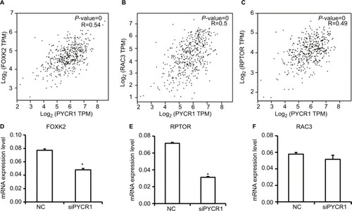
Figure 5 Analysis and detection of PYCR1 coexpression genes in human MM.
Notes: (A–C) PYCR1 coexpression genes were identified using the GEPIA dataset. FOXK2, RPTOR and RAC3 showed a similar expression pattern to that of PYCR1 in SKCM. (D–F) siPYCR1 inhibited FOXK2 and RPTOR expression, but not RAC3 expression in A375 cells. *P<0.05.
Abbreviations: MM, malignant melanoma; NC, negative control; GEPIA, Gene Expression Profiling Interactive Analysis; SKCM, skin cutaneous melanoma; FOXK2, forkhead box K2; PYCR1, pyrroline-5-carboxylate reductase; RPTOR, regulatory associated protein of MTOR complex 1.