Figures & data
Figure 1 TMEM17 expression in breast cancer tissues and cell lines. (A, B) Western blotting of 20 pairs of breast cancer tissue specimens showing that TMEM17 expression in breast cancer tissues was significantly higher than that in the corresponding normal breast tissues (p=0.015). (Ca, b) TMEM17 was negative in the normal breast duct glandular epithelium cells and weakly to moderately positive in the cytoplasm of normal breast duct myoepithelial cells. TMEM17 showed moderate (c, d) and strong (e, f) positive staining in the cytoplasm of cancer cells. (g, h) TMEM17 expression was significantly higher in breast cancer cells than in the adjacent normal breast duct glandular epithelium cells in the same field of view. Additionally, scattered nuclear staining was rarely visible in the cells. a, c, e, g 200×; b, d, f, h 400×. (D) TMEM17 expression was significantly higher in MCF-7, T47D, MDA-MB-231, and MDA-MB-468 breast cancer cells than in the normal human mammary epithelial cell line MCF-10A.
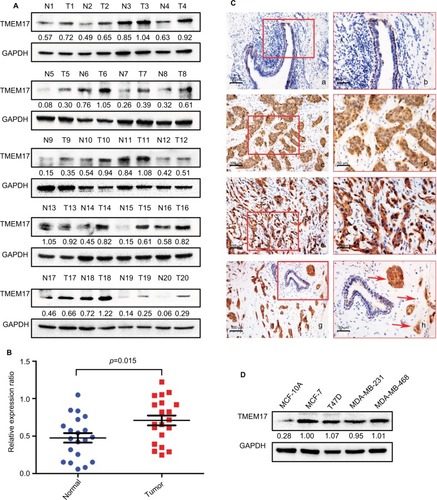
Table 1 TMEM17 expression in adjacent normal breast tissue samples and invasive breast cancer specimens
Table 2 Summary of correlation of TMEM17 expression with clinicopathological characteristics of invasive breast cancer
Figure 2 TMEM17 knockdown inhibited proliferation, colony formation, invasion, and migration of MCF-7 and MDA-MB-231 cells. (A) Interference efficiency in MCF-7 and MDA-MB-231 cell lines after TMEM17 was knocked down by siRNA. (B) MTT assay shows that TMEM17 interference significantly inhibited proliferation in MCF-7 (the fifth day, p<0.001) and MDA-MB-231 (the fifth day, p<0.001) cells. (C) Colony formation assays revealed that TMEM17 knockdown significantly decreased the number of colonies of MCF-7 (p=0.007) and MDA-MB-231 (p=0.011) cells. (D) Transwell analysis revealed that TMEM17 knockdown inhibited the invasion of MCF-7 (p=0.0025) and MDA-MB-231 (p=0.0037). (E) Wound assay revealed that TMEM17 silencing also inhibited the migration of MCF-7 (p=0.0363) and MDA-MB-231 cells (p=0.0044).
Abbreviations: siRNA, small interfering RNA; siNC, small interfering negative control.
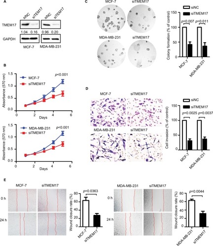
Figure 3 TMEM17 upregulated active β-catenin and Snail by affecting p-AKT and p-GSK3β, but not p-ERK. TMEM17 could not affect the expression of p-ERK or total ERK in MCF-7 and MDA-MB-231 cells. However, TMEM17 could stably upregulate p-AKT (Ser 473), p-GSK3β (Ser 9), active β-catenin, and Snail; upregulate c-myc and cyclin D1; and downregulate E-cadherin expression.
Abbreviations: siNC, small interfering negative control. NC, negative control.
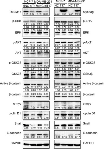
Figure 4 TMEM17 promoted the proliferation and invasion of cancer cells via activation of AKT/GSK3β signaling. (A) The AKT inhibitor LY294002 (10 μM) could inhibit the TMEM17-induced increase with p-AKT (Ser 473) and p-GSK3β (Ser 9), and downregulate active β-catenin and Snail, and then restore the expression of c-myc, cyclin D1, and E-cadherin. (B) MTT assay revealed that increased cell proliferation caused by TMEM17 was reversed by LY294002 (the fifth day, p<0.001). (C) Transwell assay revealed that the increased invasiveness caused by TMEM17 was also reversed by LY294002 (p=0.002).
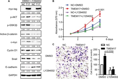
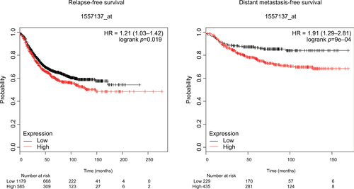
Figure S1 TMEM17 expression is significantly correlated with the relapse-free survival and distant metastasis-free survival of breast cancer patients.