
Figures & data
Table 1 Clinical characteristics of patients in the EGFR-TKI-sensitive group and nonsensitive group
Figure 1 The five types of tumor margin as seen on the CT images.
Note: (A) smooth; (B) lobulated; (C) spiculated; (D) angular; (E) obscured.
Abbreviation: CT, computed tomography.
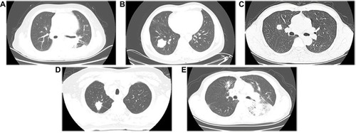
Figure 2 The four special signs seen on the CT images.
Note: (A) pleural indentation; (B) air bronchogram; (C) cavitation; (D) calcification.
Abbreviation: CT, computed tomography.
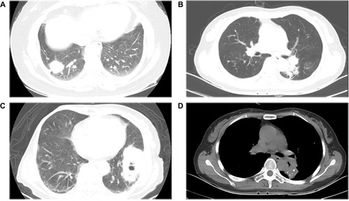
Table 2 Clinical characteristics of patients with EGFR mutation subtypes
Table 3 CT features of patients with EGFR-TKI-sensitive group and nonsensitive group
Table 4 CT features of patients with EGFR mutation subtypes
Figure 3 Relationship between tumor size and EGFR mutation subtypes.
Abbreviation: EGFR, epidermal growth factor receptor.
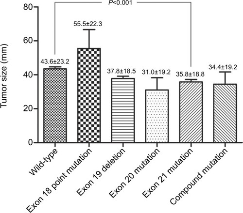
Table 5 Histological classifications in the EGFR-TKI-sensitive group and nonsensitive group