Figures & data
Table 1 Primer sequences for real-time PCR
Figure 1 Metformin induces apoptotic cell death in lung cancer cells.
Notes: (A) A549 and H460 cells were treated with metformin (20, 40, and 80 mM) for 48 hours. Cell viability was measured and quantified by MTT assay. (B) A549 and H460 cells were treated with indicated concentrations of metformin for 24 hours. Then, indicated proteins were detected by Western blot, GAPDH or tubulin was detected as an input control. (C) The cells were pretreated with z-VAD-FMK (10 µM) for 1 hour, and then exposed to metformin for 48 hours. Cell viability was measured and quantified by MTT assay. N=3. *P<0.05, ***P<0.001.
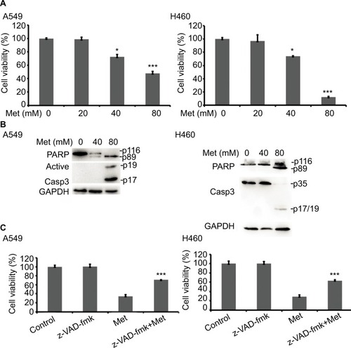
Figure 2 Metformin downregulated the expression of c-FLIPL in NSCLC.
Notes: (A) A549 and H460 cells were treated with metformin (80 mM) for 16 hours. Then, indicated proteins were detected by Western blot, and GAPDH or tubulin was detected as an input control. (B) Lung cancer cells were treated with metformin for 8 hours. Then, total RNA were extracted, and the mRNA level of c-FLIPL was examined by qRT-PCR, β-actin was detected as an input control. (C) A549 and H460 cells were pretreated with metformin (80 mM) or PBS for 4 hours, and then treated with CHX (200 µg/mL) for indicated times. Then, indicated proteins were detected by Western blot and tubulin was detected as an input control. N=3.
Abbreviations: c-FLIPL, cellular FLICE-inhibitory protein large; FLICE, FADD-like IL-1β-converting enzyme; NSCLC, non-small cell lung cancer.
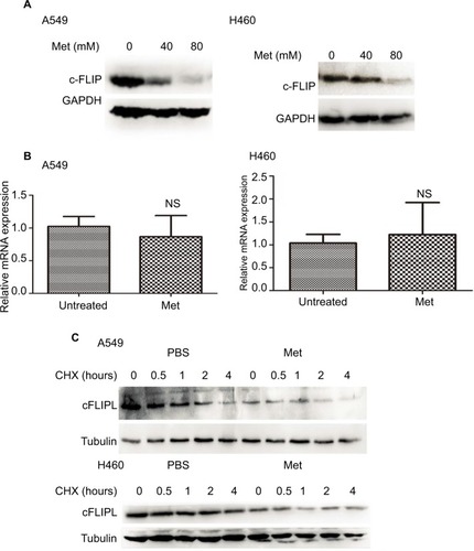
Figure 3 Metformin inhibits c-FLIPL expression through AMPK.
Notes: (A) A549 and H460 cells were treated with metformin (20, 40, 80 mM) for 2 hours. Then, indicated proteins were detected by Western blot, and tubulin was detected as an input control. (B) The cells were transfected with siRNA (10 nM) for 24 hours, and then treated with metformin (80 mM) for 24 hours. Then, cell viability was detected by MTT assay. (C) A549 and H460 cells were transfected with siRNA (10 nM) for 24 hours, and then treated with metformin (80 mM) for 16 hours. Then, indicated proteins were detected by Western blot, and tubulin was detected as an input control. N=3. **P<0.01, ***P<0.001.
Abbreviations: AMPK, adenosine 5′-monophosphate-activated protein kinase; c-FLIPL, cellular FLICE-inhibitory protein large; FLICE, FADD-like IL-1β-converting enzyme.
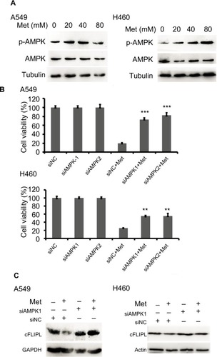
Figure 4 Metformin suppressed the expression of c-FLIPL by inhibition of PKA activity.
Notes: (A) A549 and H460 cells were treated with metformin (5, 10, 20, 40, 80 mM) for 8 hours. Then, indicated proteins were detected by Western blot, and GAPDH was detected as an input control. (B, C) Lung cancer cells were treated with 8-Br-cAMP (1 mM) and forskolin (10 µM) for 8 hours. Then, indicated proteins were detected by Western blot, and tubulin was detected as an input control.
Abbreviations: c-FLIPL, cellular FLICE-inhibitory protein large; FLICE, FADD-like IL-1β-converting enzyme; PKA, protein kinase A.
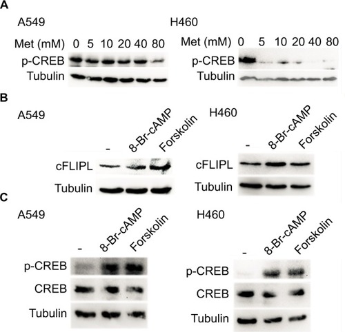
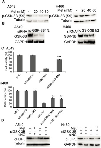
Figure 5 Metformin promotes c-FLIPL degradation via GSK-3β.
Notes: Metformin inhibits c-FLIPL expression through AMPK. (A) A549 and H460 cells were treated with metformin (20, 40, 80 mM) for 8 hours. Then, indicated proteins were detected by Western blot, and tubulin was detected as an input control. (B, C) The cells were transfected with siRNA (10 nM) for 24 hours, and then treated with metformin (80 mM) for 24 hours. Then, cell viability was detected by MTT assay. (D) A549 and H460 cells were transfected with siRNA (10 nM) for 24 hours, and then treated with metformin (80 mM) for 16 hours. Then, indicated proteins were detected by Western blot, and tubulin was detected as an input control. N=3. **P<0.01, ***P<0.001.
Abbreviations: AMPK, adenosine 5′-monophosphate-activated protein kinase; c-FLIPL, cellular FLICE-inhibitory protein large; FLICE, FADD-like IL-1β-converting enzyme; GSK-3β, glycogen synthase kinase 3 beta.