Figures & data
Table 1 Immunohistochemical staining of GIT1 in all patientderived OS tissues
Figure 1 GIT1 expression in human osteosarcoma tissues.
Notes: Two typical cases of primary osteosarcoma. In (A) there is a diffuse cytoplasmic staining, while no immunoreactivity can be detected in (B). No nuclear staining for GIT1 has been obtained (original magnification: ×400).
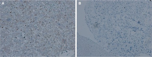
Figure 2 Knockdown efficacy of GIT1.
Notes: (A) The relative transcription of GIT1 mRNA in three kinds of osteosarcoma cell lines (HOS, 143B and OST) detected by qRT-PCR. (B) The knockdown efficacy by GIT1-shRNA in OST cells detected by real-time RT-PCR. (C) The relative expression of GIT1 protein in three kinds of osteosarcoma cell lines (HOS, 143B and OST) detected by Western Blotting. (D) The knockdown efficacy by GIT1-shRNA in OST cells detected by Western Blotting. (E) Microscopic images of OST infected with lentivirus. One-way ANOVA for comparing GIT1 mRNA among HOS, 143B and OST cells or among blank, NC-shRNA and GIT1-shRNA groups was conducted respectively (scale bar: 100 µm; *P<0.05).
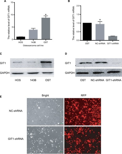
Figure 3 Knockdown of GIT1 inhibited invasion of osteosarcoma cells.
Notes: OST cells were infected with GIT1-shRNA, NC-shRNA or blank control for 24 and 48 hours respectively. Cell invasion was evaluated by the transwell migration assay. (A) The representative images of three groups. (B) Quantitative results of cell invasion assay. One-way ANOVA for comparing the number of invade cells among blank, NC-shRNA and GIT1-shRNA group was conducted (scale bar: 20 µm; *P<0.05).
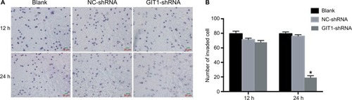
Figure 4 Knockdown of GIT1 inhibited angiogenesis of osteosarcoma.
Notes: (A) The ELISA results of VEGF concentration released from OST cells infected with GIT1-shRNA or NC-shRNA. (B) The representative images of immunohistochemistry of CD34 (a marker of tumor MVD) in vivo. (C) The statistical analysis of B. The independent Student’s t-test for comparing VEGF concentration or CD34 expression level between the two groups was conducted respectively (Scale bar: 20 µm; *P<0.05).
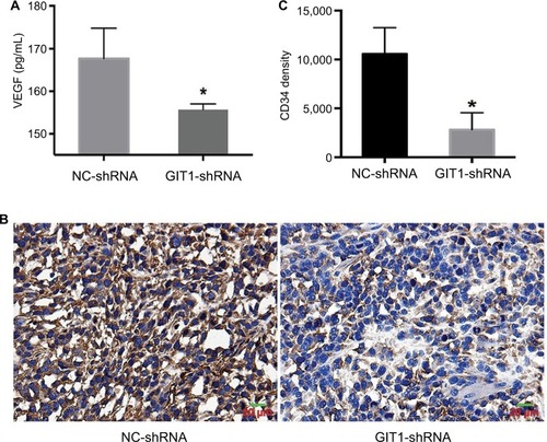
Table 2 Data of lung metastasis
Figure 5 Effect of GIT1 on primary osteosarcoma growth and lung metastasis in the orthotopic model.
Notes: OST cells infected with GIT1-shRNA or NC-shRNA were orthotopically transplanted into the distal femur of nude mice and allowed to form tumors. (A) Tumor diameter was evaluated for 45 days. (B) The representative fluorescence imaging of orthotopic tumor in two groups. The independent Student’s t-test for comparing tumor volume between NC-shRNA and GIT1-shRNA group was conducted in different post-implantation days. (C) The representative fluorescence imaging of lung metastasis in two groups. Left column: the images of whole mice; right column: the images of lung metastasis (*P<0.05).
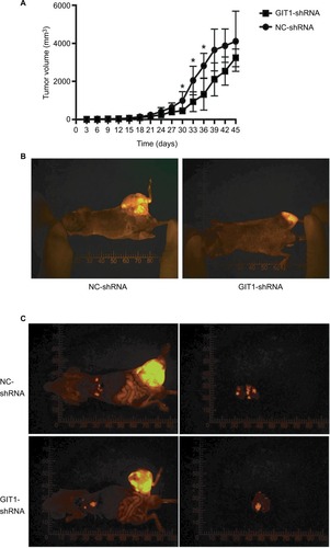
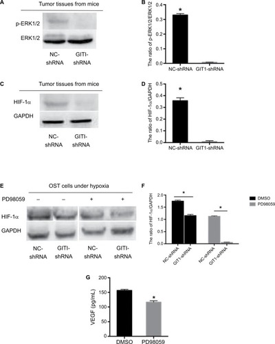
Figure 6 ERK1/2/HIF-1α pathway participated in GIT1-regulated VEGF release.
Notes: (A) The activation of ERK1/2 in tumor tissues from two groups’ mice. (B) The statistical analysis of A. (C) The expression of HIF-1α in tumor tissues from two groups` mice. (D) The statistical analysis of C. (E) In order to induce HIF-1α expression, OST cells of two groups were cultured in hypoxia for 12 hours with or without PD98059 pretreated. The expression of HIF-1α under different conditions was detected by Western blotting. (F) The statistical analysis of C. (G) The ELISA results of VEGF concentration released from OST cells infected with GIT1-shRNA with or without PD98059 pretreated. The independent Student’s t-test for comparing p-EKR1/2 or HIF-1α protein level between NC-shRNA and GIT1-shRNA group was conducted, respectively (*P<0.05).