Figures & data
Figure 1 The expression of the HER2 protein in SM-AP1, SM-AP4, and fibroblast cells.
Notes: The HER2 protein was expressed in SM-AP1 and SM-AP4 cells, but not in the fibroblasts. Magnification 400×.
Abbreviation: HER2, human epithelial growth factor receptor 2.
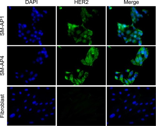
Figure 2 HER2 amplification was detected in SM-AP1 (A) and SM-AP4 (B) cells, but not in the fibroblasts (C).
Notes: Increased copy numbers (more than six) in cells are shown in A and B. HER2/CEP17 ratio =1 is shown in C. Magnification 1000×.
Abbreviation: HER2, human epithelial growth factor receptor 2.
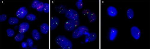
Figure 3 The effect of lapatinib on HER2 expression and its downstream pathways in SM-AP1 (A) and SM-AP4 (B) cells.
Notes: (A) p-HER2, AKT, p-AKT, and p-ERK were inhibited by lapatinib in a dose-dependent manner in SM-AP1 cells. (B) p-HER2 was inhibited by lapatinib in a dose-dependent manner, and p-ERK was suppressed significantly when the concentration of lapatinib reached at 1 µM in SM-AP4 cells.
Abbreviations: HER2, human epithelial growth factor receptor 2; p-AKT, phosphorylated AKT; p-ERK, phosphorylated ERK; p-HER2, phosphorylated HER2.
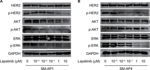
Table 1 Cell cycle analysis
Figure 4 Lapatinib inhibits the activity and proliferation of SM-AP1 (A) and SM-AP4 (B) cells, but not those of normal fibroblasts (C).
Note: *Significant at P<0.001.
Abbreviation: CON, control group.
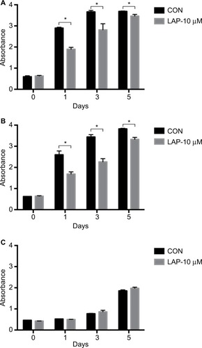
Figure 5 The effect of lapatinib on cell cycle of SM-AP1 and SM-AP4 cells.
Note: (A and B) SM-AP1 cell cycle arrested in the G1 phase after lapatinib treatment; (C and D) SM-AP4 cell cycle arrested in the G1 phase after lapatinib treatment; (E and F) normal fibroblast cell cycle was not affected by lapatinib treatment.
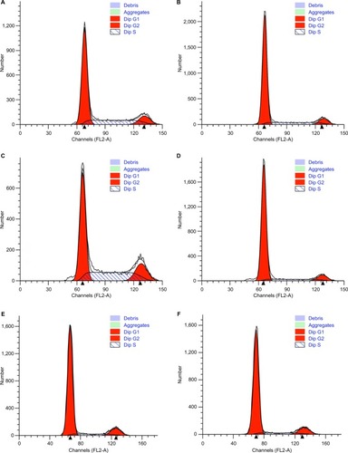
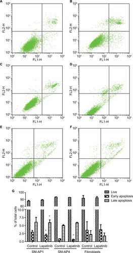
Figure 6 Lapatinib promoted the apoptosis of SM-AP1 (A and B) and SM-AP4 cells (C and D), but not that of normal fibroblasts (E and F). The percentage of apoptotic cells was increased in SM-AP1 cells, whereas the percentage of early apoptotic cells was increased in SM-AP4 cells (G). The apoptosis of fibroblasts was not affected by lapatinib (G).
Note: *Significant at P<0.001.