Figures & data
Table 1 Physico-pathological characteristics of NPC patients
Table 2 Imaging of skull base bone destruction
Table 3 The features of 17 NPC patients with nasopharyngeal hemorrhage
Table 4 Demographics and outcomes of 17 NPC patients with nasopharyngeal hemorrhage
Table 5 Clinical characteristics of 10 NPC patients with just 1 course of RT
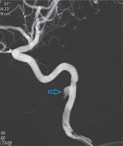
Figure 1 External carotid artery angiogram showing the bleeding sites on the internal maxillary artery (indicated by the arrow).
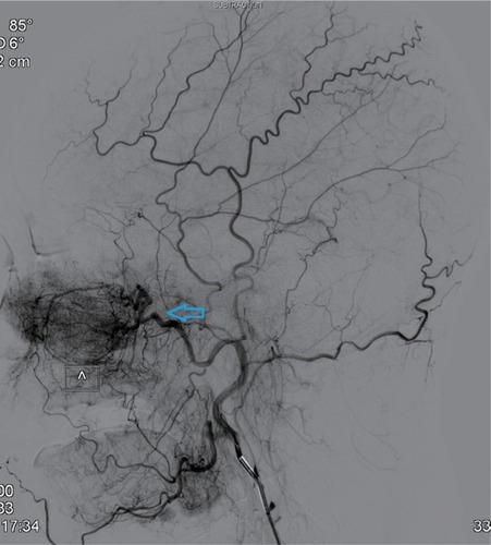
Figure 2 Post-embolization internal maxillary artery angiograms revealing total occlusion of the artery (indicated by the arrow).
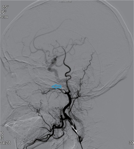
Figure 3 External carotid artery branch angiogram showing the bleeding sites on ascending pharyngeal artery (indicated by the arrow).
Note: L= blood vessel on the left.
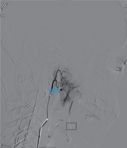
Figure 4 Post-embolization ascending pharyngeal artery angiogram revealing total occlusion of the artery (indicated by the arrow).
Note: L= blood vessel on the left.
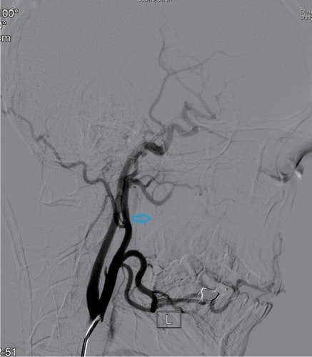
Figure 5 Right lateral internal carotid artery angiogram showing radiation arteritis with pseudoaneurysm (indicated by the arrow).
Note: RCCA=right common carotid artery.
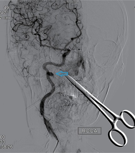
Figure 6 Vascular imaging of the pseudoaneurysm after 3D reconstruction (indicated by the arrow).