Figures & data
Figure 1 High expression of TMEFF1 correlates with worse clinical outcomes in ovarian cancer patients and the expression of TMEFF1 in ovarian cancer cell lines.
Notes: (A) Expression of TMEFF1 in various types of ovarian tissues: (a) normal ovary; (b) benign epithelial ovarian tumor; (c) borderline epithelial ovarian tumor; and (d) epithelial ovarian cancer (×400). (B) Influence of TMEFF1 expression on the OS of ovarian cancer patients. (C–E) Protein and mRNA expressions of TMEFF1 in four ovarian and one normal ovarian cell lines.
Abbreviation: OS, overall survival.
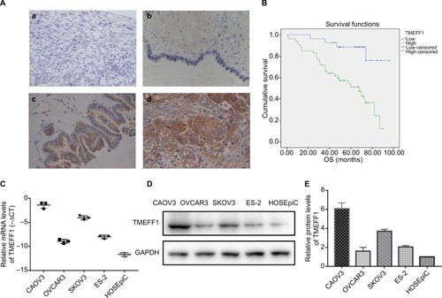
Table 1 Expression of TMEFF1 in different ovarian serous tissues
Table 2 Relationships between TMEFF1 and the clinical pathological parameters in malignant ovarian serous tumors
Table 3 Cox’s regression analysis of prognosis
Figure 2 Detection of the transfection of TMEFF1 in ovarian cancer cell lines.
Notes: (A–C) Downregulation of TMEFF1 in CAOV3 and SKOV3 ovarian cancer cell lines, overexpression of TMEFF1 in OVCAR3 and ES-2 ovarian cancer cell lines, and mRNA and protein expressions of TMEFF1. (D) The expression of TMEFF1 was detected via immunocytochemistry in ovarian cancer cells (×200 and ×400). *P<0.05, **P<0.01, and ***P<0.001.
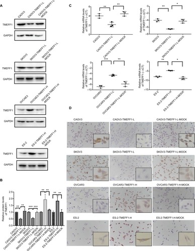
Figure 3 TMEFF1 promoted proliferation, migration, and invasion in CAOV3 and OVCAR3 ovarian cancer cell lines.
Notes: (A–C) Influence of TMEFF1 expression on the proliferation of CAOV3 and OVCAR3 cells and differential expression of PCNA. (D and E) Influence of TMEFF1 expression on the migration of CAOV3 and OVCAR3 cells. (F and G) Influence of TMEFF1 expression on invasion by cells (×400). *P<0.05, **P<0.01, and ***P<0.001.
Abbreviation: PCNA, proliferating cell nuclear antigen.
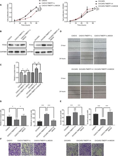
Figure 4 TMEFF1 influenced apoptosis and cell cycle in CAOV3 and OVCAR3 ovarian cancer cell lines.
Notes: (A–D) Influence of TMEFF1 expression on the apoptosis of cells and changes in bcl2/bax expression. (E and F) Influence of TMEFF1 on the cell cycle. *P<0.05, **P<0.01, and ***P<0.001.
Abbreviations: 7AAD, 7-aminoactinomycin D; PE, phycoerythrin.
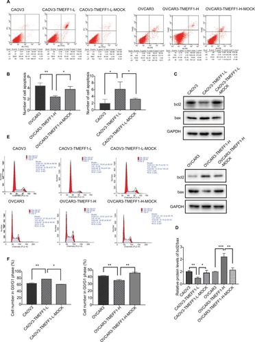
Figure 5 In CAOV3 and OVCAR3 ovarian cancer cell lines, TMEFF1 activated the MAPK and PI3K/AKT signaling pathways and regulated the expression of EMT-related proteins.
Notes: (A and B) In CAOV3 and OVCAR3 cells, TMEFF1 increased the expression of vimentin, N-cadherin, MMP2, and MMP9 but decreased the expression of E-cadherin. (C and D) Phosphorylation changes in MAPK pathway-associated nodal proteins (RAF/MEK/ERK) in CAOV3 and OVCAR3 ovarian cancer cell lines before and after TMEFF1 transfection. (E and F) Phosphorylation changes in PI3K/AKT pathway-associated nodal proteins (PI3K/AKT) after the differential expression of TMEFF1. *P<0.05, **P<0.01, and ***P<0.001.
Abbreviation: EMT, epithelial–mesenchymal transition.
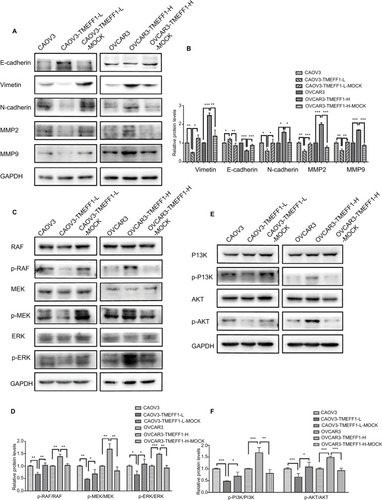
Figure 6 TMEFF1 promoted proliferation, migration, and invasion in CAOV3 and OVCAR3 ovarian cancer cell lines through MAPK and PI3K/AKT pathways.
Notes: (A) Proliferation induced by TMEFF1 in CAOV3 and OVCAR3 cells decreased after the addition of MAPK (PD98059) or PI3K (GDC-0941) pathway inhibitors. (B and C) Migration induced by TMEFF1 decreased after the addition of PD98059 or GDC-0941. (D and E) Invasion induced byTMEFF1 decreased after the addition of PD98059 or GDC-0941 (×400). *P<0.05, **P<0.01, and ***P<0.001.
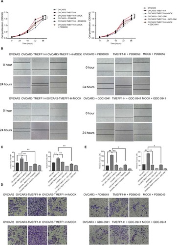
Figure 7 TMEFF1 influenced apoptosis and cell cycle in CAOV3 and OVCAR3 ovarian cancer cell lines through MAPK and PI3K/AKT pathways.
Notes: (A and B) After the addition of PD98059 or GDC-0941, the effect of TMEFF1 in decreasing the apoptosis was suppressed. (C and D) After the addition of GDC-0941, the effects of TMEFF1 in decreasing the proportion of cells in G0/G1 phase and increasing cells in S and G2–M phases were suppressed, which was not found after the addition of PD98059 (P>0.05). *P<0.05, and **P<0.01.
Abbreviations: 7AAD, 7-aminoactinomycin D; PE, phycoerythrin.
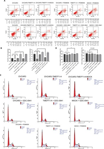
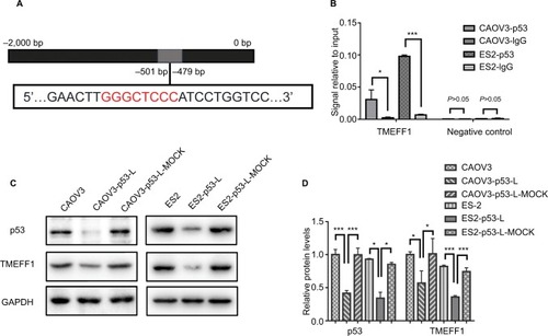
Figure 8 In ovarian cancer cells, TMEFF1 was regulated by p53.
Notes: (A) Predicted p53-binding sites in the promoter region of TMEFF1. (B) The promoter region of TMEFF1 could bind with the transcription factor, p53, in CAOV3 and ES-2 ovarian cancer cell. (C and D) The same change in TMEFF1 expression was observed with the differential expression of p53 (all P>0.05). *P<0.05, and ***P<0.001.