
Figures & data
Figure 1 A summary of proteases secretion by GBM cells and their actions upon activation.
Notes: Several positive feedback loops of activation exist. What is also evident is the extensive redundancy in the activation and upregulation of the different proteases which have overlapping functions. Research should aim to identify the factors upregulated in different subtypes of GBM to allow potential tailored treatments to different patients.
Abbreviations: ECM, extracellular matrix; GBM, glioblastoma multiforme; suPAR, soluble form of uPAR.
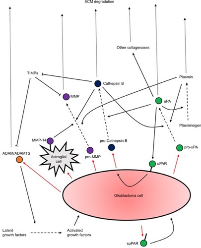
Figure 2 A Matrigel assay: an invasion assay which allows the experimenters to assess the invasive potential of cells through the Matrigel-filled pores in a membrane separating the two compartments; this resembles invasion through the ECM.
Abbreviation: ECM, extracellular matrix.
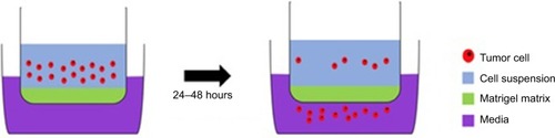
Figure 3 Diagram showing the hydrodynamic model of invasion.
Notes: Activation of GPCR (eg, by bradykinin binding to B2R) causes intracellular Ca2+ to rise, leading to Ca2+-dependent K+ and Cl− efflux through CIC3 and KCa3.1 channels. Water then follows out of the cell through aquaporins down its osmotic gradient. This decreases cellular volume, allowing the glioma cells to migrate. They then increase volume via ion influx through NKCC1 and ASIC1 channels.
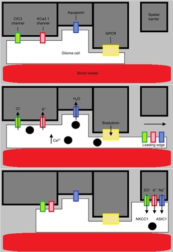
Table 1 A summary of the current ongoing Phase III and Phase IV trials