Figures & data
Figure 1 The schematic representation of the study flow.
Abbreviation: US, ultrasound.
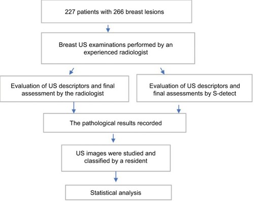
Table 1 The pathological types of the 266 breast lesions
Figure 2 The ROC for the experienced radiologists, S-detect, and the in-training resident.
Abbreviation: ROCs, receiver operating characteristic curves.
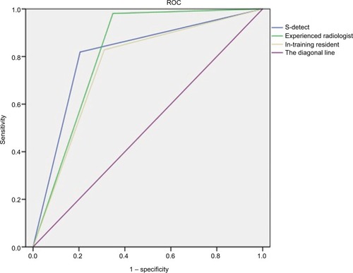
Figure 3 The breast lesion of a 51-year-old woman, without any symptoms, which proved to be ductal carcinoma in situ.
Note: (A) The longitudinal section of the lesion; (B) the cross-section of the lesion; (C) the abundant peripheral blood vessels of the tumor; and (D) the lesion was diagnosed by S-detect as a possibly benign tumor.
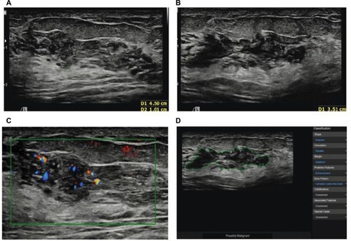
Table 2 The diagnostic performance of the experienced radiologists, S-detect, and the in-training resident
Table 3 The subcategorization of breast lesions by an experienced radiologist
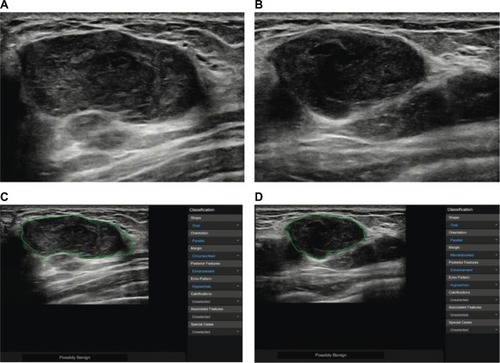
Figure 4 The breast lesion of a 46-year-old woman, without any symptoms, which proved to be chronic inflammation by histology.
Note: (A) The longitudinal section of the lesion; (B) the cross-section of the lesion; (C) the abundant peripheral blood vessels of the tumor; and (D) the lesion was diagnosed by S-detect as a possibly benign tumor.
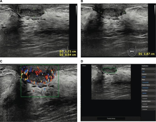
Figure 5 The breast lesion of a 35-year-old woman who found a palpable breast mass with a diameter of 2 cm 6 months ago, whereas it measured as 4 cm in this US examination. The pathological result was mucinous carcinoma.
Note: (A) The longitudinal section of the lesion; and (B) the cross-section of the lesion; (C, D) the lesion was misdiagnosed by S-detect as a possibly benign tumor.
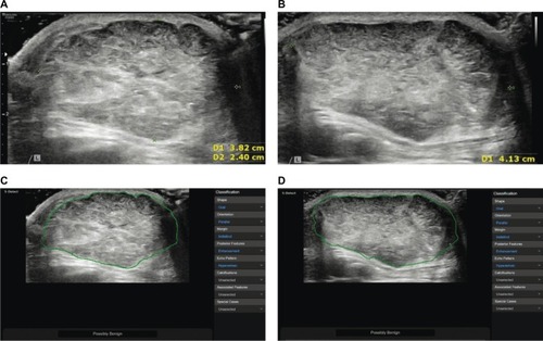
Figure 6 The breast lesion of a 41-year-old woman with a history of invasive phyllodes tumor.
Note: (A) The longitudinal section of the lesion; (B) the cross-section of the lesion; and (C, D) the lesion was misdiagnosed by S-detect as a possibly benign tumor.