Figures & data
Table 1 Associations between Wnt5a expression and clinicopathological features of esophageal squamous cell carcinoma
Figure 1 Wnt5a is highly expressed in the invasive ESCC tissues. (A) The representative images of Wnt5a expression in the pathological sections of ESCC and nonmalignant esophageal tissues. Wnt5a is highly expressed in invasive ESCC tissue. DAB substrate solution was applied to reveal the brown color of Wnt5a antibody staining. Hematoxylin staining showed the blue color of nucleus. Bar =100 μm. Objective lens, magnification, ×20; numerical aperture, 0.75. (B)Wnt5a was highly expressed in the invasive ESCC tissues (n=16) than that in the noninvasive ESCC tissues (n=6). Wnt5a immunostaining was analyzed by the evaluation of the percentage of tumor-stained cells and staining intensity, allowing assessment of an IRS. (C) Wnt5a expression was not shown correlation with the clinical and pathological characteristics of tumor size in ESCC. Tumour size, small, ≤2 cm. Large, >2 cm. (D) Western blotting revealed the endogenous Wnt5a expression in KYSE410 and KYSE520 ESCC cells. β-actin as the loading control. (E) The Wnt5a secretion was measured and the culture media of KYSE410 and KYSE520 cells by ELISA.
Abbreviations: ESCC, esophageal squamous cell carcinoma; IRC, immunoreactive score.
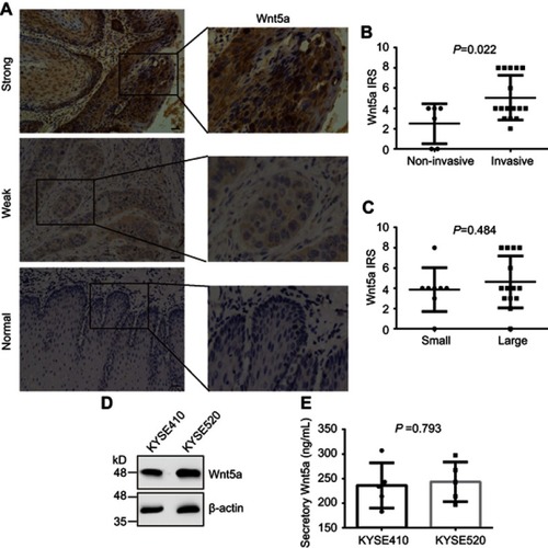
Figure 2 Wnt5a stimulates the invasion of ESCC cells. (A and B) KYSE410 and KYSE520 ESCC cells were treated with 1 μg/mL secreted Frizzled-related protein 2 (sfrp2), an antagonist of Wnt5a, and/or vehicles and allowed to invade the matrigel for 6 hrs. The invasive cells on the lower sides of Boyden chamber membranes were counted under a microscope field. Objective lens, magnification, ×20; numerical aperture, 0.75. (C and D) Wnt5a did not alter the ability of the colony formation of KYSE410 and KYSE520 cells. The single-cell suspensions of KYSE410 and KYSE520 cells were seeded on 6-well plates and were treated with 1 μg/mL sfrp2 and/or vehicles. They were allowed to grow in a normal cell-cultural condition for 15 days. Tumor cell colonies were stained with crystal violet and counted the number.
Abbreviation: ESCC, esophageal squamous cell carcinoma.
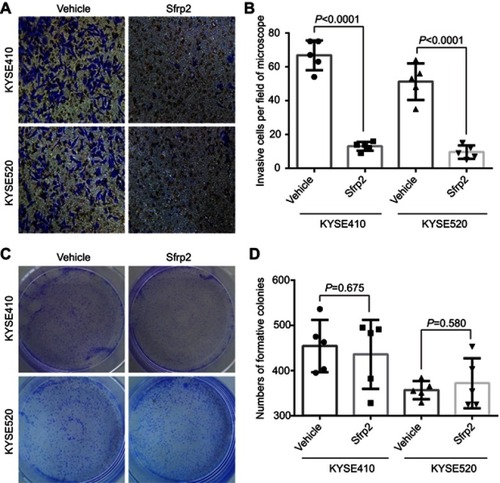
Figure 3 ROR1 and ROR2 receptors are required for the invasion of ESCC cells. (A) KYSE410 and KYSE520 were transfected with scrambled shRNA, shRNA against ROR2 (ROR2-shRNA), or ROR2-shRNA plus shRNA-resistant WT ROR2 and then subjected to the Western blotting assays. β-actin as the loading control. (B and C) Cell invasion rate was largely abolished by anti-ROR1 mAb, ROR2-shRNA, or ROR2-shRNA plus shRNA-resistant WT ROR2. KYSE410 (B) and KYSE520 (C) cells were incubated with anti-ROR1 mAb, transfected with ROR2-shRNA or ROR2-shRNA plus shRNA-resistant WT ROR2, and then subjected to cell invasion assays. (D)The lysate of KYSE410 cells was subjected to immunoprecipitation (IP) with antibody to ROR2, followed by immunoblotting with antibody to ROR1 or ROR2. (E) ROR1 was directly binding to ROR2. Purified GST or GST-ROR1 was incubated with purified Flag-tagged full-length ROR2. The amounts of Flag-ROR2 co-purified with GST or GST-ROR1 (pull down) were analyzed by immunoblotting for anti-Flag or anti-GST.
Abbreviations: ESCC, esophageal squamous cell carcinoma; WT, wild type; ROR, receptor tyrosine kinase-like orphan receptor.
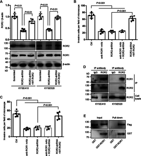
Figure 4 RhoA activation is regulated by ROR1/ROR2 and required for ESCC cell invasion. (A) KYSE410 cells were treated with anti-ROR1 mAb or vehicle and then measured the activation of Rac1, Rac2, Rac3, RhoA, and Cdc42 by G-LISA assays. (B) KYSE410 cells were transfected with ROR2-shRNA or scrambled shRNA and then measured the activation of Rac1, Rac2, and RhoA by G-LISA assays. (C) ROR2-knockdown KYSE410 and KYSE520 cells by shRNA transfection were treated with anti-ROR1 mAb, or rescued by transfected with shRNA-resistant WT-ROR2. The lysates of these cells were assayed for the evaluation of RhoA activation by G-LISA assays. (D) KYSE410 and KYSE520 cells were seeded on the Boyden chambers and treated with CCG-1423 (RhoA-specific inhibitor, 1 μmol/L) or vehicle for 1 hr, and then subjected to cell invasion assays.
Abbreviations: ESCC, esophageal squamous cell carcinoma; ROR, receptor tyrosine kinase-like orphan receptor.
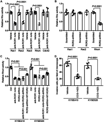
Figure 5 DAAM1 acts as the upstream of RhoA and mediates cell invasion. (A) KYSE410 and KYSE520 cells were transfected with DAAM1-shRNA or scrambled shRNA and then examined the RhoA activity by G-LISA assays. (B) DAAM1 activation was not altered by RhoA-specific inhibitor CCG-1423 treatment. KYSE410 and KYSE520 cells were treated with 1 μmol/L CCG-1423 or vehicle for 1 hr. Cellular lysates were assayed for the active DAAM1 by a pulldown assay using a GST-RhoA as a bait. (C) DAAM1 acted as the downstream target of ROR1/ROR2. KYSE410 and KYSE520 cells were treated with anti-ROR1 mAb or transfected with ROR2-shRNA. Cellular lysates were assayed for the active DAAM1 by a pulldown assay using a GST-RhoA as a bait. (D) KYSE410 and KYSE520 cells were transfected with DAAM1-shRNA and rescued by shRNA-resistant wildtype (WT) DAAM1 overexpression, and then subjected to invasion assays.
Abbreviations: ROR, receptor tyrosine kinase-like orphan receptor; WT, wild type.
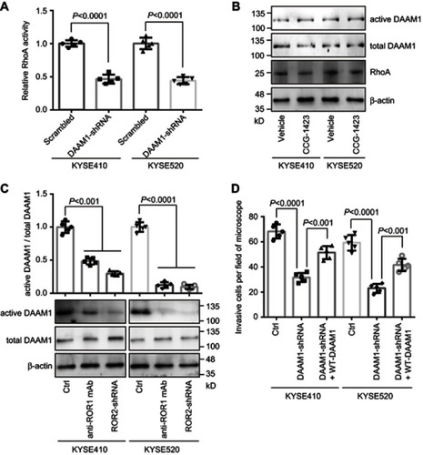
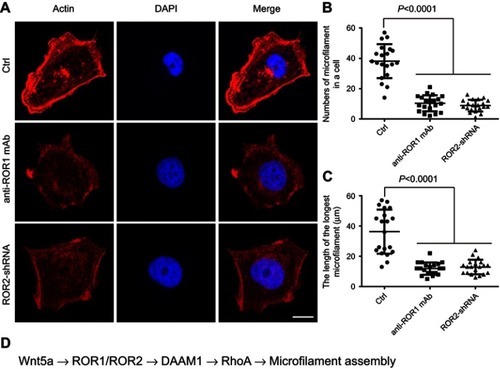
Figure 6 ROR1 and ROR2 participate in the rearrangement of microfilaments. (A) Blocking ROR1/ROR2 signaling by ROR2-shRNA transfection or anti-ROR1 mAb treatment disrupted the formation of microfilaments. KYSE410 cells treated with anti-ROR1 mAb or transfected with ROR2-shRNA were grown on coverslips. Subsequently, cells were fixed and stained with phalloidin and DAPI to reveal filamentous actin (F-actin) organization and nucleus location. Bar =10 μm. Objective lens, magnification, ×40; numerical aperture, 0.95. (B and C) The number of microfilaments in a cell and the length of the longest microfilament were determined in KYSE410 cells. Date was shown in the above scatter diagram. n=20. (D) The scheme of signaling pathway involving in the invasion of ESCC cells.
Abbreviations: ESCC, esophageal squamous cell carcinoma; ROR, receptor tyrosine kinase-like orphan receptor.