Figures & data
Figure 1 Notes: (A) Expression of CEACAM19 in non-cancer penile tissues and penile cancer. Bars: 100 µm. Upper panel, low expression of CEACAM19 in non-cancer penile tissues (1–3); middle panel, penile cancer cases with over-expression of CEACAM19 (Case 1–3); lower panel, penile cancer cases with low expression of CEACAM19 (Case 4–6). (B) Over-expression of CEACAM19 in penile cancer was associated with unfavorable CSS. (C) Nodal status predicted unfavorable CSS in penile cancer. The log-rank test was used to compare survival curves.
Abbreviation: CSS, cancer-specific survival.
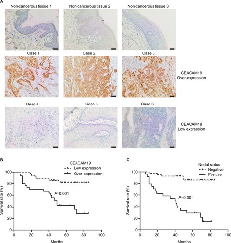
Table 1 Demographic and clinicopathological characteristic of study population associated with CEACAM19 expression
Table 2 Demographic and clinicopathological characteristic of study cohort associated with nodal status
Table 3 Cox proportional hazard model for prognosis associated with clinicopathological parameters and CEACAM19 expression
Figure 2 Knockdown of CEACAM19 expression suppresses cell growth and clonogenesis in Penl1 cells.
Notes: (A) CEACAM19 expression was significantly reduced in Penl1 cells transduced with shCEACAM19 lentivirus. β-Actin served as a loading control. (B) Knockdown of CEACAM19 expression attenuated cell growth of Penl cells. The CCK-8 absorbance was measured at 450 nm (OD450). *P<0.05, Scr vs shCEACAM19. (C) Depletion of CEACAM19 expression reduced clonogenesis of Penl1 cells. The colony formed in Scr control was regarded as 100%. *P<0.05, Scr vs shCEACAM19. All experiments were performed three times, and data are presented as mean ± SD. values. *P<0.05, Scr vs shCEACAM19.
Abbreviations: CCK-8, Cell Counting Kit-8; Scr, scramble; shCEACAM19, shRNA targeting CEACAM19.
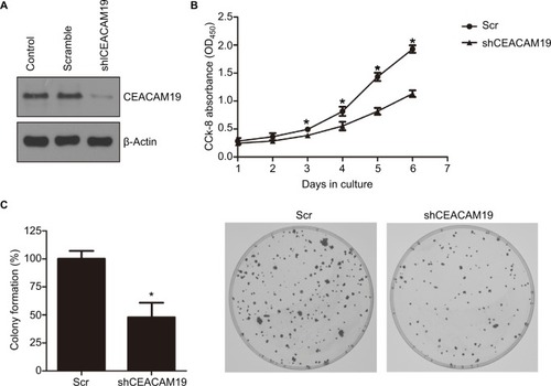
Figure 3 Depletion of CEACAM19 expression inhibits cell migration and invasion in Penl1 cells.
Notes: (A) Wound healing assay. The migration of the cells to the wound was measured at 0 and 24 hours after scratch. The representative fields were photographed; the relative healing rates were quantified with measurements of the gap sizes after the culture. Three different areas in each assay were chosen to measure the distances of migrating cells to the origin of the wound. Bars: 100 µm. *P<0.05, Scr vs shCEACAM19. (B) Transwell invasion assays with Penl1 cells were performed in Scr control and shCEACAM19 group. Crystal violet assay (OD570) was conducted to evaluate cell migration and invasion capability. Bars: 100 µm. All experiments were performed three times, and data are presented as mean ± SD. values. *P<0.05, Scr vs shCEACAM19.
Abbreviations: Scr, scramble; shCEACAM19, shRNA targeting CEACAM19.
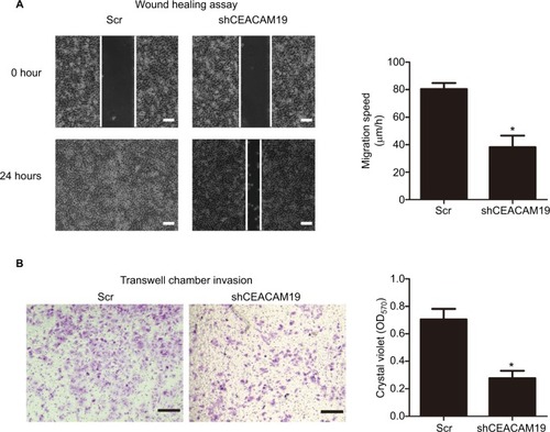
Figure 4 Notes: (A) Multiple pathway reporter analysis identified potential cancer-related pathways regulated by CEACAM19 in Penl1 cells. (B) Knockdown of CEACAM19 expression attenuated p-Smad2/3 expression in Penl1 cells. β-Actin served as a loading control. (C) Knockdown of CEACAM19 expression reduced MMP2/9 secretion in Penl1 cells. All experiments were performed three times, and data are presented as mean ± SD values. *P<0.05, Scr vs sh CEACAM19.
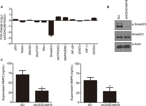
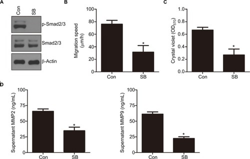
Figure 5 Notes: (A) The effect of SB431542 (SB) on p-Smad2/3 levels in Penl1 cells. Penl1 cells were treated with 10 µM SB for 24 hours. β-Actin served as a loading control. (B) The effect of SB431542 on wound healing of Penl1 cells. Three different areas in each assay were chosen to measure the distances of migrating cells to the origin of the wound. *P<0.05, Control vs SB. (C) The effect of SB431542 on cell invasion of Penl1 cells. Transwell invasion assay was conducted to evaluate cell migration and invasion capability. *P<0.05, Control vs SB. (D) Inhibition on TGF-β/Smad2/3 activity by SB431542 reduced MMP2/9 secretion in Penl1 cells. All experiments were performed three times, and data are presented as mean ± SD. values. *P<0.05, Control vs SB.