
Figures & data
Figure 1 Measurement of the distance to the papilla (DtP). The distance shown in the figure is 1.17 cm.
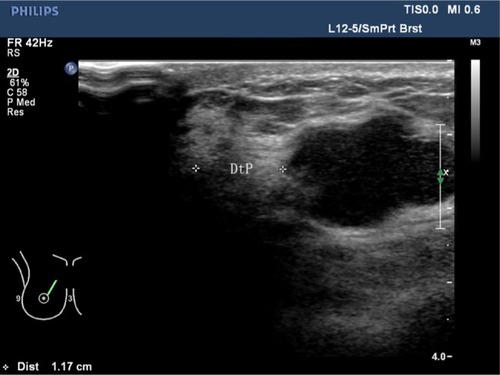
Figure 2 Measurement of the distance from the superficial edge of the lesion to the skin (DtS) and the distance from the deep edge of the lesion to the pectoralis muscle (DtPM).
Note: The distances shown in the figure are DtS =0.752 cm and DtPM =0.191 cm.
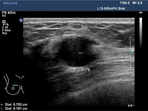
Figure 3 The benign sub-models of contrast-enhanced ultrasound for breast lesions. (A) Rapid wash-in with hyper-enhancement and clear margin after enhancement without enlarged size. (B) Synchronous or slow wash-in with iso-enhancement, and no difference in margin and shape after enhancement. (C) Synchronous or slow wash-in with hypo-enhancement.
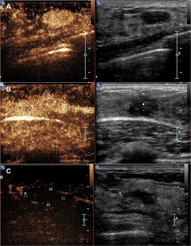
Figure 4 The malignant sub-models of contrast-enhanced ultrasound for breast lesions.
Notes: (A) Hyper-enhancement with enlarged range, with or without irregular shape. (B) Hyper-centripetal enhancement with perfusion defect, with or without enlarged range. (C) Rapid or synchronous wash-in with hyper- or iso-enhancement, presenting penetrating vessels (as indicated by the white arrows) or crab claw-like pattern, with or without perfusion defect.
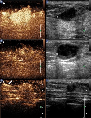
Table 1 Characteristics of the patients and lesions
Table 2 Characteristics of the patients according to the type of final diagnosis