
Figures & data
Figure 1 The cellular model of each marker positive staining.
Note: (A) Negative staining; (B) H&E staining; (C) Ki-67 nuclear positive staining; (D) p16 cytoplasmic positive staining; (E) p16 cytoplasmic and Ki-67 nuclear co-positive staining; (F) ProEx™ C nuclear positive staining; (G) HPV L1 capsid protein nuclear positive staining; (H) Claudin 1 membranous positive staining; (I) IMP3 cytoplasmic positive staining; (J) Feulgen-thionin staining for DNA; and (K) RKIP nuclear and cytoplasmic positive staining.
Abbreviations: HPV, human papillomavirus; IMP3, insulin-like growth factor-II mRNA-binding protein 3; RKIP, Raf kinase inhibitor protein.
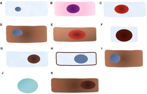
Table 1 Diagnostic performance of the p16/Ki-67 dual staining in primary screening for detecting CIN 2+
Table 2 Triage performance of the p16/Ki-67 dual staining in patients referred to colposcopy for detecting CIN 2+
Table 3 Triage performance of the p16/Ki-67 dual staining in patients with ASCUS/LSIL for detecting CIN 2+
Table 4 Triage performance of the p16/Ki-67 dual staining in patients with HPV+ for detecting CIN 2+
Table 5 Diagnostic performance of other staining marker for detecting CIN 2+