Figures & data
Figure 1 A 66-year-old woman visited our hospital due to chronic ulceration of the right foot 8 months ago. (A) There was a 3×2 cm erythematous reticular chronic ulcer lesion in the mid-plantar surface of the foot. (B) Wide excision with a margin of 1 cm or more was performed. Plantar fascia and lateral plantar neurovascular bundle were exposed through skin defect. (C) One week after the operation, a dermal matrix was formed by granulation in the skin defect. (D) Split-thickness skin graft was performed by harvesting from the ipsilateral thigh. (E) After 8 weeks postsurgery, complete healing of the skin graft was confirmed.
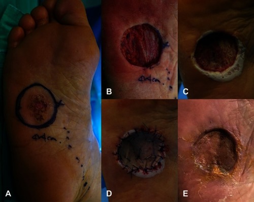
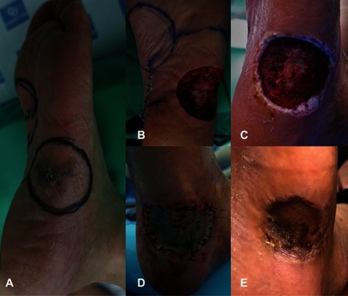
Figure 2 A 69-year-old male presented with color change in the left foot 4 months ago. (A) A 3×2 cm pigmented skin lesion was observed on the lateral side of the mid-plantar foot. (B) Wide excision with a margin of 1 cm or more was performed, and plantar fascia was exposed through skin defect. (C) One week after surgery, a dermal matrix was formed. (D) Split-thickness skin graft was performed by harvesting from the ipsilateral thigh. (E) Six weeks after the surgery, complete healing of the skin graft was confirmed.