Figures & data
Figure 1 Immunohistochemical staining of HMGA2, E-cadherin and vimentin in normal pancreatic tissue and pancreatic cancer samples. (A) No expression of HMGA2 in the normal pancreatic tissue. (B) Positive signals of HMGA2 in pancreatic cancer. (C) No expression of vimentin in the normal pancreatic tissue. (D) Positive signals of vimentin in pancreatic cancer. (E) Positive signals of E-cadherin in the normal pancreatic tissue. (F): No expression of E-cadherin in pancreatic cancer. Scale bar=50 um.
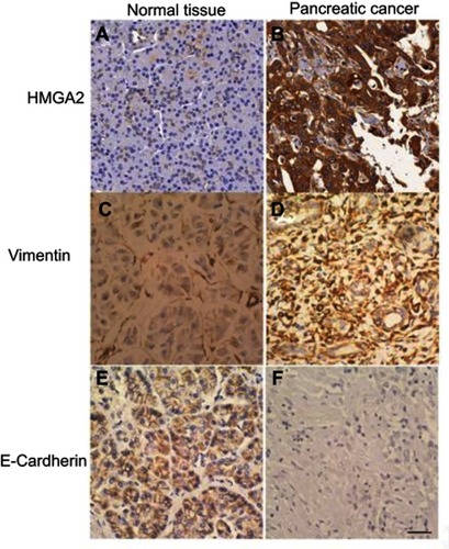
Table 1 Association of HMGA2, E-cardherin and Vimentin expression with the clinicopathological features of 60 patients with pancreatic cancer
Figure 2 Survival curves of patients with HMGA2, E-cadherin and vimentin expression in pancreatic cancer. (A) Patients with high HMGA2 levels showed significantly worse survival than those with low expression (P=0.022). (B) Patients with high vimentin levels showed significantly worse survival than those with low expression (P=0.028). (C) Patients with low E-cadherin levels had significantly worse survival than those with high expression (P=0.003).
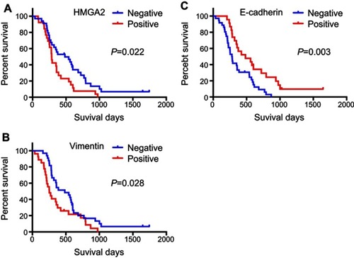
Table 2 Univariate and multivariate analysis of factors associated with overall- survival of patients with pancreatic cancer
Figure 3 HMGA2 knockdown in pancreatic cancer CAPAN 1 cells using SiRNAs (HMGA2-S1 and HMGA2-S2). (A) Quantitative real-time PCR and (B) Western blot data showing decreased HMGA2 mRNA and protein expression levels in both knockdown groups. ***P<0.001. HMGA2 over expressed in PC cells using plasmid pcDNA HMGA2 and non-target controls (pcDNA). (C) Quantitative real-time PCR and (D) Western blot data showing increased HMGA2 mRNA and protein expression levels in pcDNA HMGA2 group. **P<0.01.
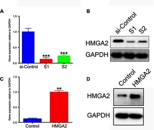
Figure 4 (A) Compared with control cells, migration and invasion abilities in HMGA2-S1 and HMGA2-S2 cells were significantly inhibited (x100). (B) Columns show the means of three independent experiments. *P<0.05, ***P<0.001. (C) Compared with control cells, migration and invasion abilities in HMGA2 overexpressed cells were significantly enhanced (x100). (B) Columns show the means of three independent experiments. *P<0.05, **P<0.01.
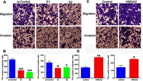
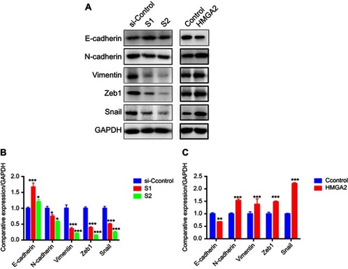
Figure 5 (A) Western blot for detecting EMT-associated protein markers in HMGA2 knockdown and overexpressed CAPAN 1 cells. (B) and (C) Semiquantitative histograms of Western blot results. *P<0.05, **P<0.01, ***P<0.001.