Figures & data
Figure 1 BCL11A mRNA overexpression level in breast cancer. (A) The relative BCL11A expression level was detected in 20 pairs of breast cancer samples. (B) qRT-PCR analysis of BCL11A expression in two normal immortalized breast cell lines (184A1 and MCF-10A) and breast cancer cell lines. **p<0.01; *p<0.05. (C) HE-staining and immunohistochemical-staining of BCL11A protein in a clinical sample and its adjacent normal tissue. Original magnification, 100× and 400×.
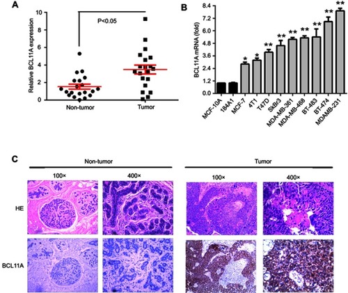
Figure 2 BCL11A has strong oncogenic functions. (A) A Western blot assay was used to characterize the expression of BCL11A in BCL11A-overexpressing and control vector-transfected cells. shRNA against BCL11A effectively decreased BCL11A expression detected by Western blotting (left). BCL11A expression was confirmed by qRT-PCR (right), and β-actin was used as a loading control. (B) MTT assays were performed to compare the cell growth rates between BCL11A-overexpressing and control cells and between BCL11A-silenced and control cells. (C) Representative images of the increased foci formation ability induced by BCL11A in MCF-7 and MDAMB231 cell lines. Quantitative analyses of foci numbers are shown in the right panel. (D) Representative images of xenografts and a summary of tumor weight in nude mice. The weights of xenograft tumors are summarized in the right panel. All results are expressed as the mean ± SD of three independent experiments, *p<0.05; **p<0.01.
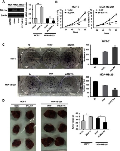
Figure 3 BCL11A promotes tumor metastasis, cancer cell migration, and the stemness of breast cancer cells. BCL11A promotes cell metastasis ability, confirmed by Transwell invasion assay in vitro. (A) Representative invaded cells image (Left), and the statistical analysis of the invaded cell number (Right) are shown. All the data was shown as the mean ± SD, and three independent experiments were performed (**p<0.01; *p<0.05). (B) Representative image of lung metastasis nodules on the lung of xenograft model (arrows). 8 weeks after tail vein injection of BCL11A-modulating MDA-MB-231 and MCF-7 cells, the number of lung nodules on lungs surface of nude mice was counted and analysed (N=3) (**p<0.01; *p<0.05). (C) Hematoxylin and eosin stained metastatic nodules on the surface of the lung. Representative image of sections was shown. Original magnification, 100×. (D) BCL11A increases the sphere-forming ability of MDA-MB-231 and MCF-7 cells. Original magnification: ×100. (E) Representative dot plots of CD44+/CD24− cell surface markers from mammospheres in BCL11A-osverexpressing MCF-7 and sh-BCL11A MDA-MB-231 and their controls.
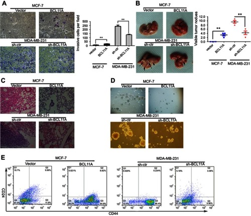
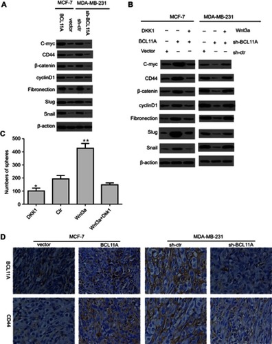
Figure 4 BCL11A promotes the breast cancer cell stemness and epithelial-mesenchymal transition by activating the Wnt/b-catenin pathway. (A) Relative C-myc, fibronectin, β-catenin, CD44, cyclinD1, slug, and snail expression level between BCL11A-modulating cells and controls of MDA-MB-231 and MCF7 was characterized by Western blotting. (B) The C-myc, CD44, fibronectin, β-catenin, cyclinD1, slug, and snail expression level was effectively decreased by Dkk1, a Wnt inhibitor, in BCL11A-overexpressing MCF-7 cells, whereas effectively increased by Wnt3a, a Wnt agonist, in MDA-MB-231 cells transfected with shBCL11A. (C) Wnt3a significantly promoted the formation of more spheres, whereas the Wnt inhibitor DKK1 reversed this trend and reduced tumor spheres number. All the data was shown as the mean ± SD, and three independent experiments were performed (**p<0.01; *p<0.05). (D) BCL11A and CD44 expression were further confirmed in xenograft tumors of nude mice through immunohistochemistry, and representative images were shown. Original magnification, 200×.