Figures & data
Table 1 Clinical characteristics of NPC patients according to high and low ZNF488 expression
Figure 1 High expression of ZNF488 is correlated with poor clinical outcomes. (A) Negative ZNF488 staining in nasopharyngeal epithelium tissue, (B) negative ZNF488 staining in NPC tissue with normal rabbit IgG, (C) negative staining of ZNF488, (D) weak staining, (E) moderate staining, (F) strong staining (magnification 400×). (G) Overall survival, (H) locoregional recurrence-free survival. (I) Distant-metastasis-free survival. (J) Progression-free survival.
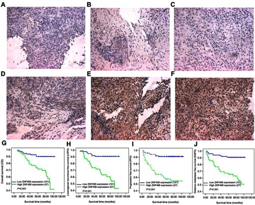
Table 2 Univariate analysis and multivariable cox regression analyses of ZNF488 expression levels and overall survival
Figure 2 Effects of ZNF488 on adhesion ability and FAK signaling pathway. (A) Immunofluoresce for ZNF488 detection. (B) Western blot to detect ZNF488 protein level in both HNE1 and CNE1 ZNF488 overexpression stable cell lines and vector. (C) Adhesion assay in HNE1 and CNE1. (D) FAK activation assay to detect the activity of pFAK (Y397). (E) Western blot to detect collagen IV, integrin α5, FAK and p-FAK(Y397). (F) Western blot to detect ERK 1/2, p-ERK1/2, Akt and p-Akt. *** P<0.001.
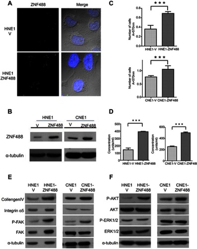
Figure 3 Effects of ZNF488 on cell proliferation. (A) The growth curves of MTT assays in both HNE1 and CNE1. (B) Colony formation assays were conducted. * P<0.05; *** P<0.001.
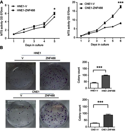
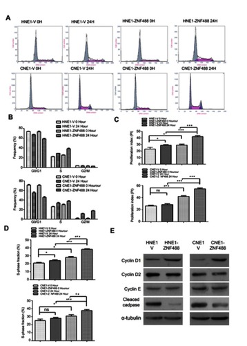
Figure 4 Effect of ZNF488 on cell cycle distribution and Cyclin D1. (A) Cell cycle analysis with flow cytometry were performed in HNE1 and CNE1. (B) Results of cell cycle analysis with histogram. (C) Proliferation index of each group. (D) S-phase fraction of each group. (E) Western blot to detect the protein level of cyclin D1, Cyclin D2, Cyclin E (C-19), and cleaved caspase 9 p10. NS, no significance. * P<0.05, ** P<0.01, *** P<0.001.