Figures & data
Table 1 Correlation between FGF5 expression and clinical-pathological characteristic in 15 osteosarcoma patients
Figure 1 (A) Heatmap of differentially expressed genes between osteosarcoma tissues and tissues adjacent to cancer. (B and C) Expression levels of FGF5 in osteosarcoma cell lines and tissues detected by qRT-PCR, the results showed that FGF5 mRNA expression was significantly increased in osteosarcoma cell lines and tissues. (D) ICH staining for FGF5 in osteosarcoma and matched normal tissues. (E and F) Protein expression levels of FGF5 in osteosarcoma cell lines and tissues detected by Western blot, the results showed that FGF5 protein expression was upregulated in osteosarcoma cell lines and tissues. Error bars indicated mean ± SD (*P<0.01).
Abbreviations: HOB, human normal osteoblast; FGF5, fibroblast growth factor-5; GAPDH, glyceraldehyde 3-phosphate dehydrogenase.
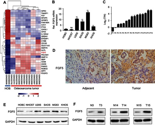
Figure 2 (A) The expression of FGF5 in two osteosarcoma cell lines after FGF5 knockout by Crispr/Cas9, the results showed that FGF5 was dramatically decreased in FGF5-KO MG63 an U20S cells. (B and C) Effects of FGF5 on cell proliferation and colony formation assessed by CCK-8 and colony formation assays. The results showed that knockout of FGF5 inhibited cell proliferation. (D) Effects of FGF5 on tumor growth determined by nude mouse orthotopic metastatic model. (E) ICH staining for FGF5 in nude mice. Error bars indicated mean ± SD (*P<0.05, **P<0.01).
Abbreviations: Crl, control; KO, knockout; FGF5, fibroblast growth factor-5; GAPDH, glyceraldehyde 3-phosphate dehydrogenase.
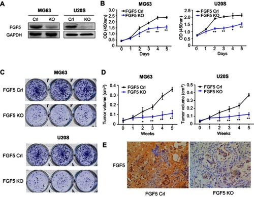
Figure 3 (A) The expression of FGF5 and FGFR1 in osteosarcoma cell after addition of exogenous rFGF5 into KHOS cell line. (B) The effect of rFGF5 on cell proliferation detected by CCK-8 assay. The results showed that exogenous rFGF5 enhanced cell proliferation. (C) The effect of rFGF5 on apoptosis detected by flow cytometry analysis and the protein expression of cleaved PARP. The results showed that exogenous rFGF5 inhibited cell apoptosis. Error bars indicated mean ±SD (*P<0.05, **P<0.01).
Abbreviations: Crl, control; FGF5, fibroblast growth factor-5; FGFR1, FGF receptor-1; rFGF5, recombinant FGF5; GAPDH, glyceraldehyde 3-phosphate dehydrogenase; CCK-8, cell counting kit-8; PARP, poly (adenosine diphosphate) ribose polymerase.
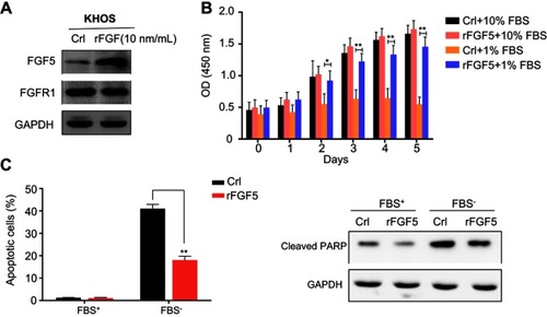
Figure 4 The expression level of FGF5, FGFR1 and MAPK signaling pathway associated proteins (p-MEK2, MEK2, p-ERK, ERK, p-MNK1, and MNK1) on control or FGF5 knockout OS cells without or with rFGF5 treatment and detected by Western blot. The results showed that FGF5 activiated the MAPK signaling. *** P <0.001 vs. Crl; ### P <0.001 vs rFGF5-
Abbreviations: Crl, control; KO, knockout; FGF5, fibroblast growth factor-5; FGFR1, FGF receptor-1; rFGF5, recombinant FGF5; OS, osteosarcoma; MAPK, mitogen-activated protein kinase; GAPDH, glyceraldehyde 3-phosphate dehydrogenase.
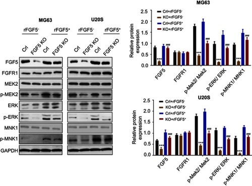
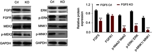
Figure 5 The expression levels of FGF5, FGFR1 and MAPK signaling pathway associated proteins (p-MEK2, MEK2, p-ERK, ERK, p-MNK1, and MNK1) in control and FGF5 knockout nude mice detected by Western blot. The results showed that FGF5 knockout dampen MAPK signaling in vivo. *** P <0.001
Abbreviations: Crl, control; KO, knockout; FGF5, fibroblast growth factor-5; FGFR1, FGF receptor-1; MAPK, mitogen-activated protein kinase; MAPK, mitogen-activated protein kinase; GAPDH, glyceraldehyde 3-phosphate dehydrogenase.