
Figures & data
Table 1 Clinicopathologic characteristics of 96 patients with sinonasal squamous cell carcinoma
Table 2 Correlation of PD-L1, p16, CD8, and Foxp3 with clinicopathologic parameters
Figure 1 Representative images of immunohistochemical staining of SNSCC tissue: (A) PD-L1-positive (100×); (B) p16-positive (100×); (C) low CD8 infiltration (400×); (D) high CD8 infiltration (400×); (E) low Foxp3 infiltration (400×); (F) high Foxp3 infiltration (400×). Abbreviations: SNSCC, sinonasal squamous cell carcinoma; PD-L1, programmed death-ligand 1.
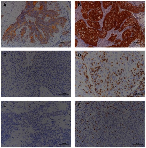
Figure 2 Box plots presenting tumor-infiltrating CD8+ and Foxp3+ cells from different groups of SNSCC: (A) CD8+-infiltrating lymphocytes and (B) Foxp3+-infiltrating lymphocytes, according to PD-L1 status; (C) CD8+-infiltrating lymphocytes and (D) Foxp3+-infiltrating lymphocytes, according to p16 status. Abbreviations: SNSCC, sinonasal squamous cell carcinoma; PD-L1, programmed death-ligand 1.
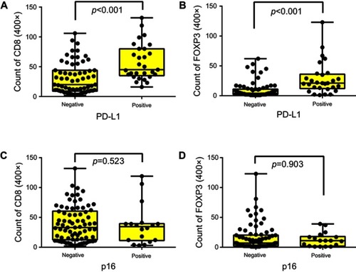
Figure 3 Correlation between Foxp3+-infiltrating lymphocytes and CD8+-infiltrating lymphocytes in SNSCC. Abbreviation: SNSCC, sinonasal squamous cell carcinoma.
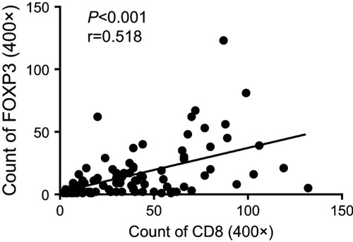
Table 3 Univariate analysis of clinicopathological features with overall survival, and disease-free survival in sinonasal squamous cell carcinoma
Figure 4 Kaplan–Meier curves for (A) OS and (B) DFS in PD-L1-negative and PD-L1-positive patients; Kaplan–Meier curves for (C) OS and (D) DFS in p16-negative and p16-positive patients; Kaplan–Meier curves for (E) OS and (F) DFS in patients stratified for high or low numbers of tumor-infiltrating CD8+ T-cells; Kaplan–Meier curves of (G) OS and (H) DFS for patients stratified for high and low numbers of tumor-infiltrating Foxp3+ T-cells.
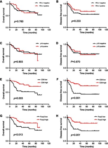
Table 4 Multivariate analysis of clinicopathological features with overall survival and disease-free survival in sinonasal squamous cell carcinoma