Figures & data
Figure 1 Structure of dydio.
Abbreviation: dydio, dihydrodiosgenin.
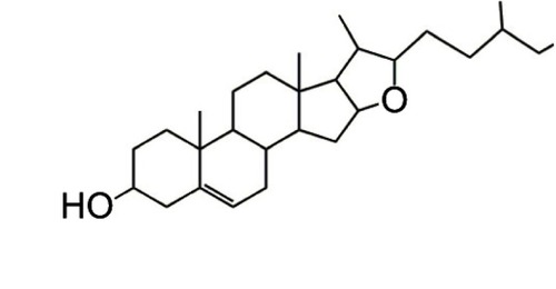
Figure 2 Effect of dydio on HCC viability and TCIPA. (A) Effect of increasing concentration of dydio on HepG2 or Hepal-6 cell viability. (B) 35 μmol/L dydio on HepG2 or Hepal-6 cell colony formation. (C) Effect of dydio on platelet aggregation induced by Hepal-6 tumor cells. (D) Effect of dydio on morphology change of platelet induced by Hepal-6 tumor cells (magnification ×100).*p<0.05, **p<0.01 vs 0 μmol/L or TC group.
Abbreviations: Ctr, control; dydio, dihydrodiosgenin; TC, tumor cells; HCC, hepatocellular carcinoma; TCIPA, tumor cell-induced platelet activation.
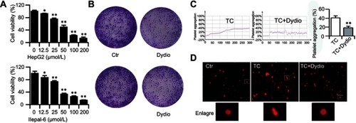
Figure 3 Dydio treatment inhibits platelet-mediated tumor cells migration. (A) Representative epifluorescence microcopy images of dil-labeled platelets (red) adhering to tumor cells (magnification ×100). (B) Platelets were pretreated with 0 or 35 μmol/L dydio, washed, and activated with Hepal-6 tumor cells to generate releasates. (C) Cell viability was assessed after 24 hours of exposure to platelet releasates. (D) Tumor cell migration was assessed after 24 hours of exposure to platelet releasates (magnification ×100). (E) E-cadherin expression was detected after 24 hours of exposure to platelet releasates. (F) Phase-contrast micrographs of Hepal-6 cell after 24 hours of exposure to platelet releasates (magnification ×200).**p<0.01 vs Plt releasates group.
Abbreviations: Ctr, control; dydio, dihydrodiosgenin; TC, tumor cells; Plt, platelets.
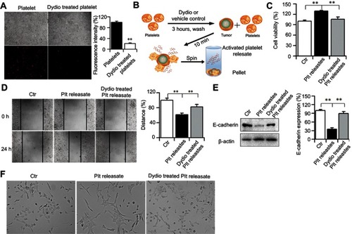
Figure 4 Dydio treatment abrogates platelet, tumor cell and ECs interaction via decreasing EC-derived FVIII release. (A) Dydio decreases the expression of FVIII in HUVEC. (B) Dydio reduce the FVIII secretion. (C) Schematic of dydio treatment on ECs. ECs were pretreated with 35 μmol/L dydio or vehicle control, washed, and then tumor cells or platelets were added and incubated for 3 hours in the static condition, washed to observe cells adhesion. (D) Representative epifluorescence microcopy images of dio–labeled HCC (green; left, HepG2; right, Hepal-6) and dil-labeled platelets (red) adhering to HUVEC (magnification ×100). **p<0.01 vs 0 μmol/L or Ctr CM.
Abbreviations: Ctr, control; dydio, dihydrodiosgenin; TC, tumor cells; Plt, platelets; CM, culture media; HUVEC, human umbilical vein cell; ECs, endothelial cells; FVIII, factor VIII.
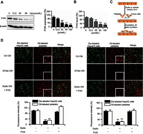
Figure 5 FVIII promotes HCC proliferation. (A) Schematic overview of the role of FVIII in dydio-mediated tumor progression. (B and C) Cell viability of HepG2 or Hepal-6 was detected after 24-hour treatment with increasing concentrations of FVIII (0, 0.25, 0.5, 1, 2, 4 and 8 U/mL). (D) Number of DNA cell cycle rate in Hepal-6 cells were analyzed by using flow cytometry.*p < 0.05, **p < 0.01 vs 0 μmol/L or Ctr group.
Abbreviations: Ctr, control; HCC, hepatocellular carcinoma; dydio, dihydrodiosgenin; FVIII, factor VIII; Dip G1, diploid first gap; Dip S, diploid synthesis; Dip G2, diploid second gap.
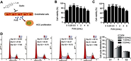
Figure 6 Dydio inhibits hepal-6 tumor cells lung metastasis highly correlated with the altered platelet function and coagulation level. (A) Representative photographs obtained from lungs of Hepal-6-injected mice. (B) The weight of lungs in each group. (C) H&E staining for lung tissues demonstrated tumor-induced thrombi formation within lung tissue. The stars indicate the tumor-induced thrombi. (D) Quantification of thrombi formation within lung tissue. (E) Platelets aggregation induced by ADP in vivo. (F) Platelets count in each group. (G) Blood clotting time in each group. (H) FVIII activities in each group.*p<0.05, **p<0.01 vs Model group.
Abbreviations: Ctr, control; dydio, dihydrodiosgenin; FVIII, factor VIII.
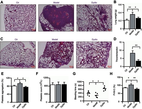
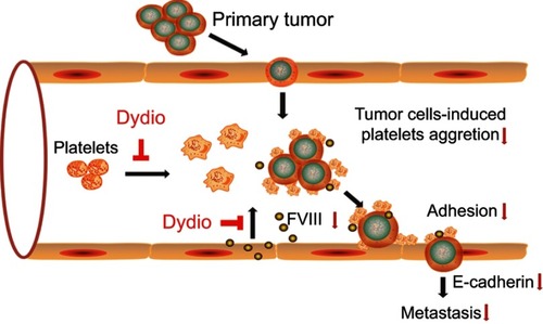
Figure 7 Schematic overview of the role of dydio in tumor metastasis.
Abbreviations: dydio, dihydrodiosgenin; FVIII, factor VIII.