Figures & data
Figure 1 Berberine suppresses the invasion, migration and proliferation of NSCLC cells and induces apoptosis in vitro. (A) Berberine inhibits A549 and PC9 cell proliferation. (B) Berberine inhibits colony formation of A549 and PC9 cells. (C) Berberine inhibits A549 and PC9 cell migration. (D) Berberine inhibits A549 and PC9 cell invasion. (E) Flow cytometry was used to quantify early and late apoptosis in A549 and PC9 cells in various concentrations of berberine for 48 h. (F) Western blot analysis was performed to examine the effects of berberine on the expression of apoptosis-related proteins. Mean values of three independent experiments are shown. Values are the mean ± SD, *P<0.01; **P<0.001, compared to vehicle controls (0.008% DMSO).
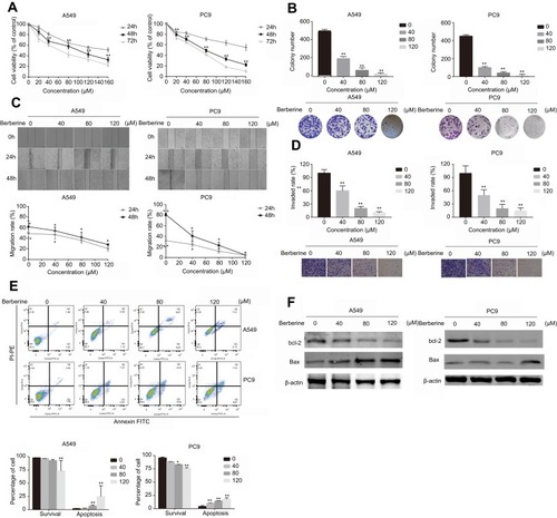
Figure 2 Berberine inhibits the growth of NSCLC and promotes apoptosis in vivo. (A) Berberine inhibits NSCLC growth in vivo. (B) Tumor weights in berberine and control groups. (D) Weights of mice in berberine and control groups. (E-F) Berberine had inhibitory effects on solid tumor cell proliferation shown by immunohistochemistry of Ki67, bcl-2. Values in each group are expressed as mean ± SD. **P<0.001.
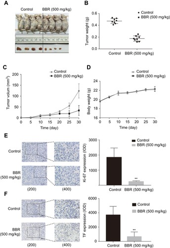
Figure 3 Berberine induces apoptosis by regulating miR-19a in NSCLC. (A) Berberine up-regulates miR19a expression in A549 and Pc9 cells in a dose-dependent manner. (B) qRT-PCR analysis of the miR-19a expression levels in miR-NC and miR-19b inhibitor transfection groups treated with berberine (80μM) for 48 h. (C) Percentage of relative cell viability after berberine treatment following transfection of the miR-19a inhibitor for 48 h. (D) Suppression of miR-19a by berberine treatment results in a significant increase in apoptotic cells. (E) Western blot analysis was performed to demonstrates that berberine induces apoptosis through miR-19a. Mean values of at least three independent experiments are shown. Values are expressed as mean ± SD, *P<0.05; **P<0.001, compared to vehicle controls (0.008% DMSO).
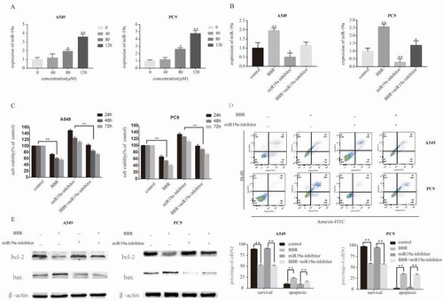
Figure 4 MiR-19a directly targets TF in NSCLC. (A) MiR-19a directly targets TF in the miRBase. (B) Luciferase activity after transfection of F3-WT or F3-MUT with miR-19a-3p mimics or mimics-NC. (C) qRT-PCR analysis of the relative expression of TF in A549 and PC9 cells transfected with miR-19a mimics or miR-19a inhibitors, with or without berberine treatment. (D) Western blot analysis of the relative TF expression in A549 and PC9 cells transfecred with miR-19a mimics or miR-19a inhibitors, with or without berberine treatment. β-Actin was used as a loading control. Values are expressed as mean ± SD, *P<0.05; **P<0.001, compared to vehicle controls (0.008% DMSO).
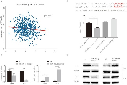
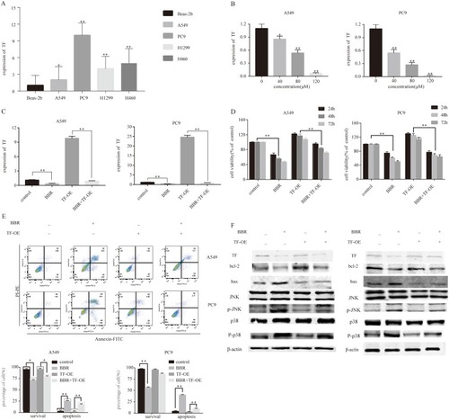
Figure 5 Berberine induces apoptosis by regulating the miR-19a/TF/MAPK axis in NSCLC. (A) qRT-PCR analysis of TF expression in BEAS-2B, A549, Pc9, H1299 and H460 cell lines. (B) Berberine up-regulates TF expression in A549 and Pc9 cells in a dose-dependent manner. (C) qRT-PCR analysis of TF expression in control and TF overexpression groups ± berberine (80 μM) for 48 h. (D) Percentage of relative cell viability after berberine treatment and TF overexpression for 48 h. (E) Suppression of TF overexpression by berberine significantly increases apoptosis. (F) Western blot analysis was performed to assess the effects of miR-19a on the expression of apoptosis-related proteins in response to berberine. Mean values of at least three independent experiments are shown. Values are expressed as mean ± SD, *P<0.05; **P<0.001, compared with the vehicle control (0.008% DMSO).