
Figures & data
Figure 1 Breast digital tomosynthesis projection acquisition (CC-view): the X-ray source rotates around the static and compressed breast over a limited angular range, while the detector is static or rotates slightly.
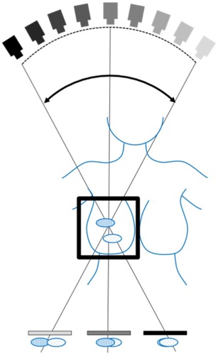
Figure 2 The strategy and scope of this review, the number of identified articles and the number considered eligible for inclusion in this work. The decision taken at each step of the eligibility process is shown in a red box.
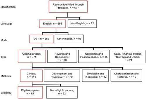
Table 1 Sensitivity, specificity and AUC - comparison of DBT alone to DM
Table 2 Recall rate and cancer detection rate - Comparison of DBT alone to DM
Table 3 Sensitivity, specificity and AUC - comparison of DM&DBT to DM alone, for all lesion types
Table 4 Recall rate and cancer detection rate - comparison of DBT&DM to DM
Table 5 Sensitivity, specificity and AUC - comparison of 1vDBT with/without 1vDM or 2vDM and/or 1vSM, to 2vDM
Table 6 Recall rate and cancer detection rate - comparison of 1vDBT with/without 1vDM or 2vDM and/or 1vSM, to 2vDM
Table 7 Sensitivity, specificity and AUC - comparison of DBT&SM or SM to DM or of SM to DM
Table 8 Recall rate and cancer detection rate - comparison of DBT&SM or SM to DM or of SM to DM