Figures & data
Figure 1 A 66-year-old male patient who was diagnosed with moderately differentiated HCC according to the pathologic examination received combined US/CT-guided RFA after TACE treatment. (A) A CT scan which was performed after TACE treatment showed the HCC lesion with intense lipiodol accumulation (arrow). (B) The HCC lesion (arrow) was incompletely visible on US because of the obstruction of air in the lung. The electrode needle (arrowhead) was inserted into the site closest to the tumor under US guidance. (C) A three-dimensional image of CT scan was used to observe the position of the probe (arrow: tumor; arrowhead: multiple electrode needle). (D) A contrast-enhanced CT scan, that was performed seven months after the combined US/CT-guided RFA procedure, showed the ablated tumor (arrow) with intense lipiodol accumulation and no local recurrence.
Abbreviations: CT, computed tomography; US, ultrasound; RFA, radiofrequency ablation; TACE, transarterial chemoembolization; HCC, hepatocellular carcinoma.
![Figure 1 A 66-year-old male patient who was diagnosed with moderately differentiated HCC according to the pathologic examination received combined US/CT-guided RFA after TACE treatment. (A) A CT scan which was performed after TACE treatment showed the HCC lesion with intense lipiodol accumulation (arrow). (B) The HCC lesion (arrow) was incompletely visible on US because of the obstruction of air in the lung. The electrode needle (arrowhead) was inserted into the site closest to the tumor under US guidance. (C) A three-dimensional image of CT scan was used to observe the position of the probe (arrow: tumor; arrowhead: multiple electrode needle). (D) A contrast-enhanced CT scan, that was performed seven months after the combined US/CT-guided RFA procedure, showed the ablated tumor (arrow) with intense lipiodol accumulation and no local recurrence.Abbreviations: CT, computed tomography; US, ultrasound; RFA, radiofrequency ablation; TACE, transarterial chemoembolization; HCC, hepatocellular carcinoma.](/cms/asset/44bd7fd7-c623-46b9-a333-fc7f96e3485b/dcmr_a_12186645_f0001_b.jpg)
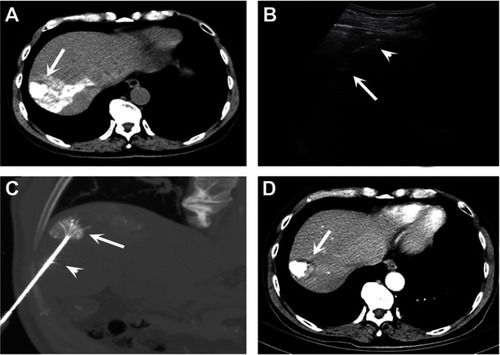
Table 1 The baseline patient characteristics