Figures & data
Figure 1 Influence of AMPH-1 on osteosarcoma progression. (A) Immunohistochemical results showing AMPH-1 protein expression in osteosarcoma and osteofibrous dysplasia tissue samples. (B, C) Western blotting analysis of AMPH-1 protein expression in 143B cells and U-2 OS cells transfected with scrambled siRNA (si N and si AMPH-1). (D, E) Cell colony formation and statistical analysis for 143B cells and U-2 OS cells transfected with scrambled siRNA (si N and si AMPH-1). (F) Flow cytometry analysis of cell apoptosis for 143B and U-2 OS osteosarcoma cells; FL1-H is annexin V-FITC and FL2-H is PI. (G) Cell cycle distribution and proportion analysis for the AMPH-1 knockdown (sh AMPH-1) and control (sh NC) groups of 143B and U-2 OS cells.
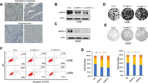
Figure 2 Influence of AMPH-1 knockdown on osteosarcoma cells. (A, B) 143B and U-2 OS cells were treated with scrambled siRNA (si N and si AMPH-1) for 72 h and then collected for Western blotting analysis. The total protein from (A) 143B and (B) U-2 OS cells was loaded and analyzed by immunoblotting with anti-p-MEK and anti-p-ERK antibodies. (C) Comparison of tumor size three weeks after inoculation of nude mice with shAMPH-1 and shNC 143B osteosarcoma cells. (D, E) Knockdown of AMPH-1 significantly increased the final tumor weights and every group involved five tumors. (F). Immunostaining of AMPH-1 and P-ERK in tumor samples.
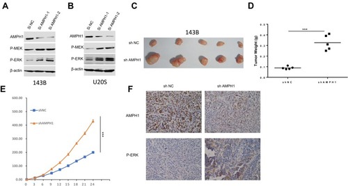
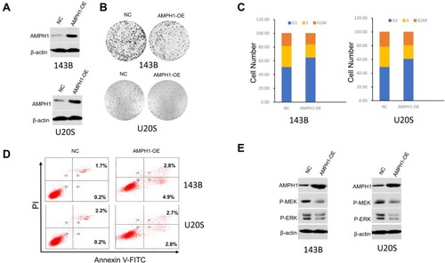
Figure 3 Inhibition of osteosarcoma growth and cell cycle arrest and promotion of cell apoptosis by overexpression of AMPH-1. (A) Western blotting analysis of AMPH-1 protein expression in 143B and U-2 OS cells transfected with AMPH-1 expression vector (AMPH-1 OE). (B) Cell colony formation by the transfected 143B and U-2 OS cells. (C) Cell cycle distribution and proportion analysis for the transfected 143B and U-2 OS cells. (D) Flow cytometry analysis of cell apoptosis for the transfected 143B and U-2 OS cells; FL1-H is annexin V-FITC and FL2-H is PI. (E) 143B and U-2 OS cells were treated with AMPH-1 expression vector (AMPH-1 OE) for 24 h and then collected for Western blotting analysis. The total protein was analyzed by immunoblotting with anti-p-MEK and anti-p-ERK antibodies.