Figures & data
Figure 1 Isolation and identification of colorectal cancer (CRC) stem cells in the SW620 cell line and determination of miR-194 and slingshot 2 (SSH2) expression levels in CRC stem cells and non-stem cells. (A) The proportions of CD44+ and CD133+ cells were analyzed by flow cytometry before and after isolation with microbeads. Before isolation, CD44+/CD133+ cells accounted for 4.53% of the total number of cells. After isolation, CD44+/CD133+ cells accounted for 88.7% of the total number of cells. (B) CRC stem cells were cultured in serum-free medium and sphere formation was evaluated under a microscope after 10 days. Scale bar = 100 µm. (C) RT-qPCR results showed that the expression levels of miR-194 were reduced (*P<0.05 according to the two-tailed t test). (D)The expression levels of SSH2 were increased in CRC stem cells compared with those in CRC non-stem cells (*P<0.05 according to the two-tailed t test). (E) The SSH2 protein expression levels were increased in CRC stem cells compared with those in CRC non-stem cells.
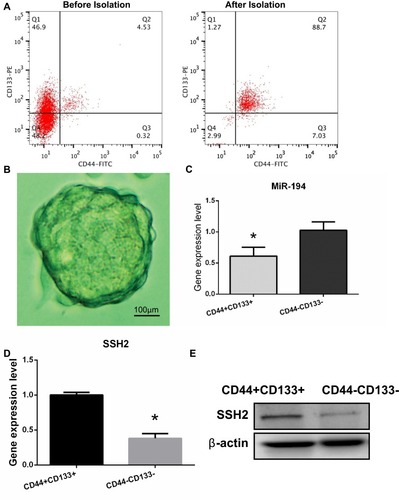
Figure 2 miR-194 inhibits cell proliferation, blocks the G1/S transition, and promotes apoptosis in colorectal cancer (CRC) stem cells by directly targeting the slingshot 2 (SSH2) gene. (A) The 3′-UTR of SSH2 contains three binding sites for miR-194. (B) NC represents CRC stem cells transfected with miR-194-NC; miR-194 represents CRC stem cells transfected with miR-194. The SSH2 mRNA expression levels, determined via quantitative RT-PCR, were reduced in CRC stem cells transfected with miR-194 compared with those in CRC stem cells transfected with miR-194-NC (*P<0.05). (C) The SSH2 protein expression level, determined via Western blot, was reduced in CRC stem cells transfected with miR-194 compared with that in CRC stem cells transfected with miRNA-194-NC. (D) Schematic presentation of luciferase reporter vector, this containing either the wild-type or mutant 3′-UTR of SSH2 were co-transfected into HEK 293T cells with miR-194-NC or miR-194. (E) Compared with co-transfection with miR-194-NC, co-transfection with miR-194 led to a significant decrease in the luciferase activity of the pGL3-SSH2-3′ UTR-Wt reporter (P<0.05 according to the two-tailed t test), but not in that of the pGL3-SSH2-3′-UTR-Mut reporter. (F) Colony formation assay demonstrating that CRC stem cells transfected with miR-194-NC formed a substantially higher number of colonies than CRC stem cells transfected with miR-194(P<0.05 according to the two-tailed t test). (G) MTT assay showing that CRC stem cell proliferation was significantly decreased in miR-194 transfected cells compared with that in cells transfected with miRNA-194-NC (P<0.05 according to ANOVA). (H) miR-194 blocks the G1/S transition in CRC stem cells. Cells were transfected with miR-194 or miR-194-NC and subjected to flow cytometry for cell-cycle analysis. The results showed that, compared with CRC stem cells transfected with miRNA-194-NC, the proportion of miRNA-194-transfected CRC stem cells at the G1 phase was increased, whereas the proportion at the S phase was markedly reduced(P<0.05 according to the two-tailed t test). (I) The effect of miR-194 and miR-194-NC on CRC stem cell apoptosis was investigated by Annexin V–FITC/PI staining. A greater percentage of apoptotic cells were observed among miR-194-transfected cells than among those transfected with miRNA-194-NC(P<0.05 according to the two-tailed t-test).
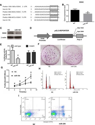
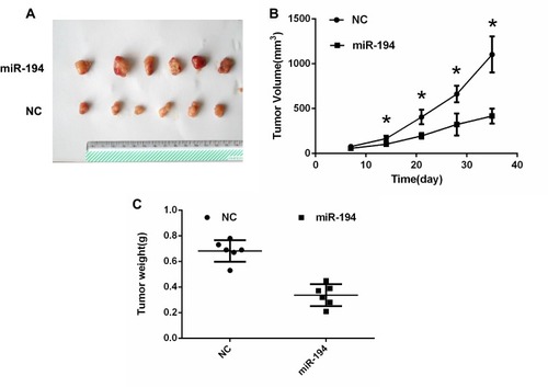
Figure 3 Colorectal cancer (CRC) stem cells transfected with miR-194 or miR-194-NC were implanted subcutaneously into the shoulder area of mice. (A) A xenograft tumor model was established in nude mice. At 14 days, The tumors were removed from the animal models and displayed. (B)Tumor growth with implantation of CRC stem cells transfected with miR-194 was considerably slower than that with implantation of CRC stem cells transfected with miR-194-NC at 14,21,28,35 days after inoculation (*P < 0.05 according to ANOVA; n = 6). (C) At 5 weeks after implantation, tumor weight with implantation of CRC stem cells transfected with miR-194 was lower than that with implantation of CRC stem cells transfected with miR-194-NC (P<0.05 according to the two-tailed t-test; n = 6).