Figures & data
Table 1 Antibody Composition of Three Panels for Differentiating Lymphocyte Subsets
Figure 1 Gating strategy of T cell subsets (Panel 1). Gating the lymphocytes by physical characteristics (A). T cells were identified by CD3 staining (B), and TCR αβ, TCR γδ were gated from T cells (C). CD4 and CD8 stainings were used to gate Th, Tc (D), Effector memory subsets of Th and Tc can be further divided into CD45RA and CD197 (E, F).
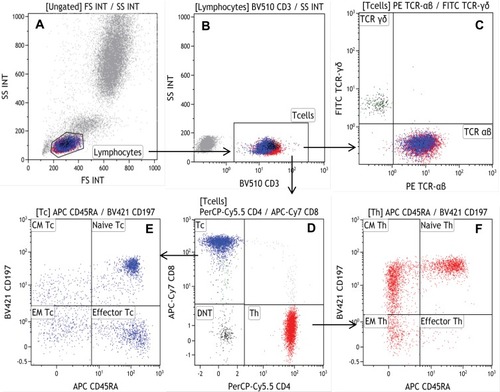
Figure 2 Gating strategy of NKT and T, NK cell subsets (Panel 2). Lymphocytes were gated according to their size and granularity in forward (FS INT)/side scatter (SS INT) (A). Treg can be identified with CD25 expression and low or negative expression of CD127 (C). Th1, Th2 and Th17 were gated from Th (B) that can be identified by CD196 and CD183 expression (D), the difference between NK and NKT cells was identified whether there was CD3 expression (E), the NK subpopulation can be divided with CD16 staining (F), CD3-CD56-CD16+ cell population (G) may be associated with HCV infection or AIDS (autoimmune diseases).
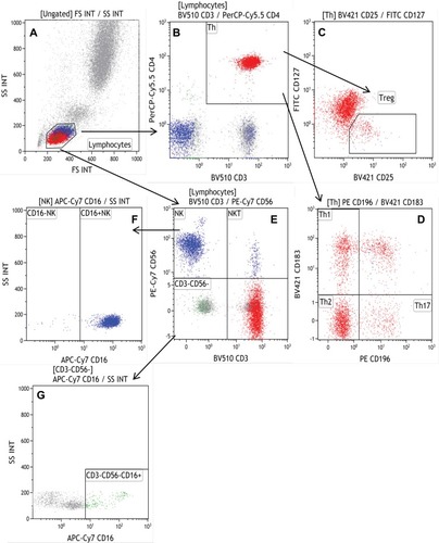
Figure 3 Gating strategy of B cell subsets (Panel 3). B cells were separated from lymphocytes (A) by CD19 staining (B), and CD27 versus CD38 gating (C) allowed the separation of B cells, including Breg stained by CD24 (D), and without CD5 and CD24 expression on plasma blasts (E); however, CD5 and CD38 were expressed on translational B cells (F), Naïve B cells identified by negative expressions of CD27, CD38, CD5 (G), and CD24 and CD27 were expressed on memory B cells (H).
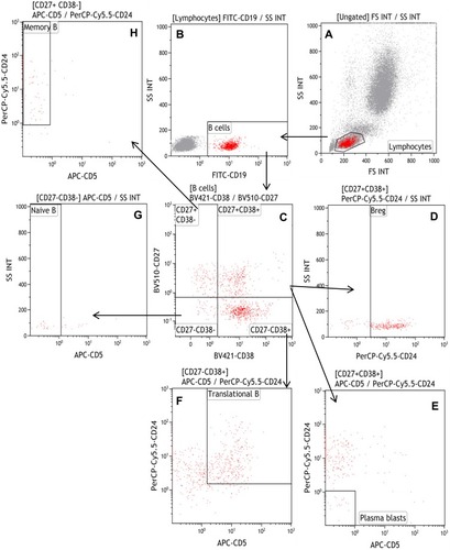
Table 2 Reference Intervals of Lymphocyte Subsets and Indicators
Table 3 The Correlation of Lymphocyte Subsets Between Males and Females by T-Test Analyses
Table 4 Differences in Lymphocyte Subsets Between Patients with Malignant Solid Tumors and Healthy Donors
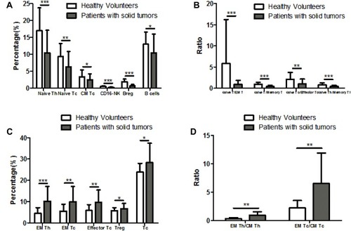
Figure 4 Differences in Lymphocyte subsets between patients with malignant solid tumors and healthy donors. There were decreased percentages or ratios of Naïve Th, Naïve Tc, CM Tc, CD16-NK, Breg, B cells (A) and Naïve T/EM T, Naïve T/Memory T, Naïve Tc/Effector Tc, Naïve Th/Naïve Tc (B) in patients with solid tumors compared with healthy donors. However, there were increased percentages or ratios of EM Th, EM Tc, Effector Tc, Treg, Tc (C) and EM Th/CM Th, EM Tc/CM Tc (D) in patients with solid tumors compared to healthy donors. *P <0.05, **P <0.01, ***P <0.001.