Figures & data
Figure 1 Emodin inhibits phosphorylation of Stat3 in pancreatic cancer cells. The PANC-1 and BxPC-3 cells were incubated with emodin at the concentration of 30, 60 or 90 μM. p-Stat3 expression in the cells was detected by Western blotting.
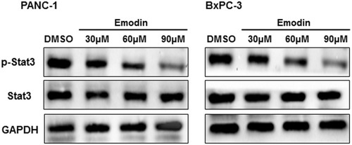
Figure 2 Emodin promotes apoptosis of pancreatic cancer cells. The PANC-1 and BxPC-3 cells were incubated with emodin at the concentration of 30, 60 or 90 μM. (A) Flow cytometry was used to detect the apoptosis of cells, and the right is the quantitative analysis of flow cytometry data. (B) Bcl-2, cleaved-caspase3, and Bax expressions in the cells were detected with Western blotting. *P<0.05; **P<0.01.
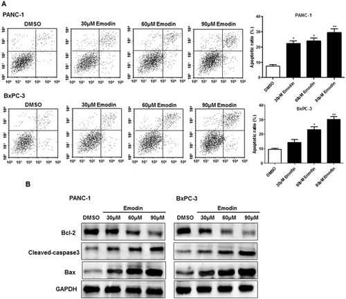
Figure 3 The overexpression of Stat3 promotes proliferation of pancreatic cancer cells. (A) The mRNA and (B) protein expression of Stat3 were detected with qRT-PCR and Western blotting, respectively. (C) The proliferation of cells was detected with MTT assay.
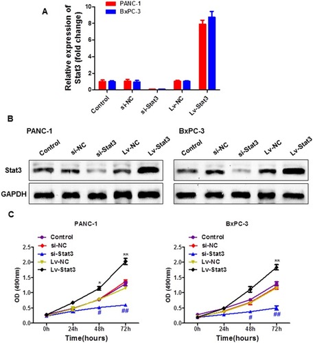
Figure 4 EGFR inhibitor promotes phosphorylation of Stat3 in pancreatic cancer cells. The EGFR inhibitor (erlotinib, gefitinib, afatinib and cetuximab) was used to treat the PANC-1 and BxPC-3 cells at the concentration of 20 nM. (A) The mRNA and (B) protein expressions of EGFR were detected with qRT-PCR and Western blotting, respectively. (C) And the protein expression of p-Stat3 was detected with Western blotting.
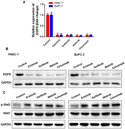
Figure 5 Emodin combined with EGFR inhibitor inhibits proliferation of pancreatic cancer cells in vitro. (A) The colon and (B) MTT assays were used to detect the proliferation in PANC-1 and BxPC-3 cells.
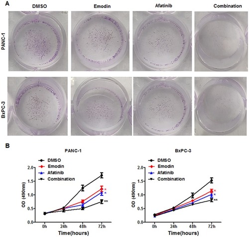
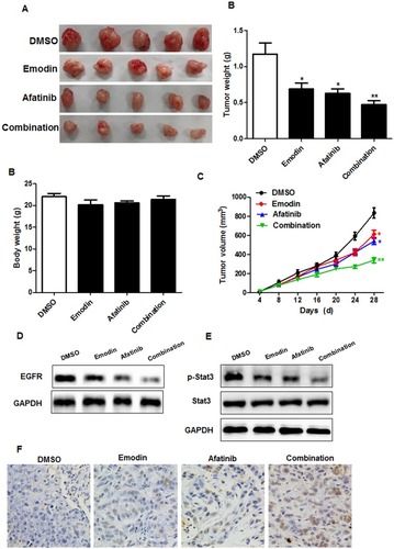
Figure 6 Emodin combined with EGFR inhibitor inhibits proliferation of pancreatic cancer cells in vivo. Mice were administrated with vehicle (saline), emodin at 50 mg/kg/day, afatinib at 50 mg/kg/day, or the combination of the 2 drugs for 4 weeks. Tumor size and tumor weight (A), body weight (B) and tumor volume (C) of mice were measured. The protein of EGFR (D) and p-Stat3 (E) were detected with Western blotting. TUNEL assay detected the apoptosis in the tumor tissues (F). *P<0.05; **P<0.01.