
Figures & data
Figure 1 PHD2 and HIF-1α mRNA expression in normal endometrium, atypical endometrial hyperplasia, and endometrial cancer. (A) PHD2 expression was reduced in endometrial cancer compared with normal endometrium (p<0.05, chi-square test). (B) The expression of HIF-1α was elevated in endometrial carcinoma compared with normal endometrium (p<0.05, chi-square test).
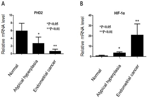
Figure 2 Western blot analysis of PHD2 and HIF-1α protein expression in normal endometrium, atypical endometrial hyperplasia, and endometrial cancer. (A) PHD2 expression was reduced in endometrial cancer compared with normal endometrium (p<0.05, chi-square test). (B) HIF-1α expression was elevated in endometrial cancer compared with normal endometrium (p<0.05, chi-square test). Each protein sample analysis was repeated in triplicate.
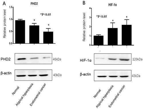
Table 1 PHD2 Staining in Endometrial Tissues
Figure 3 Immunohistochemical staining of PHD2 in endometrial tissues. (A) High expression of PHD2 in normal endometrium (×400); (B) moderate expression of PHD2 in atypical endometrial hyperplasia (×400); (C) low expression of PHD2 in endometrial cancer (×400); (D) low expression of HIF-1α in normal endometrium (×400); (E) moderate expression of HIF-1α in atypical endometrial hyperplasia (×400); (F) high expression of HIF-1α in endometrial cancer (×400).
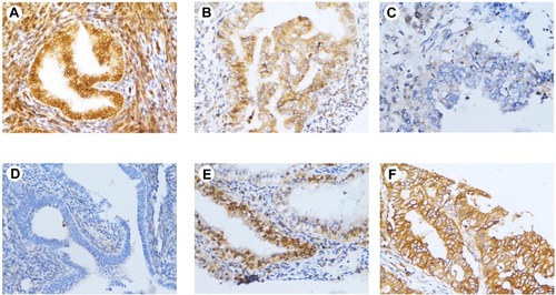
Table 2 Relationship of PHD2 and HIF-1α Expression with Clinicopathologic Characteristics of EC