
Figures & data
Figure 1 Sevoflurane suppresses cell viability, increases caspase-3 activity and up-regulates miR-203 expression in osteosarcoma cells. U2OS and MG63 cells were exposed to different concentrations of sevoflurane (1%, 2%, 5% and 10%) for 6 hrs (A and B) MTT assay determined the cell viability of U2OS and MG63 cells; (C-D) caspase-3 activity assay kit determined the caspase-3 activity of U2OS and MG63 cells; (E-F) qRT-PCR determined the expression of miR-203 in U2OS and MG63 cells. N = 3. *P<0.05, **P<0.01 and ***P<0.001.
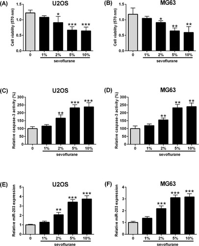
Figure 2 MiR-203 overexpression suppresses cell viability, increases caspase-3 activity and inhibits cell invasion of osteosarcoma cells. U2OS and MG63 cells were transfected with mimics NC, miR mimics or untreated, and 24 hrs later, (A, B) qRT-PCR determined the expression of miR-203 in U2OS and MG63 cells; (C, D) MTT assay determined the cell viability of U2OS and MG63 cells; (E, F) caspase-3 activity assay kit determined the caspase-3 activity of U2OS and MG63 cells; (G, H) colony formation assay determined the cell growth of U2OS and MG63 cells; (I, J) Transwell invasion assay determined cell invasive ability of U2OS and MG63 cells. N = 3. *P<0.05, **P<0.01.
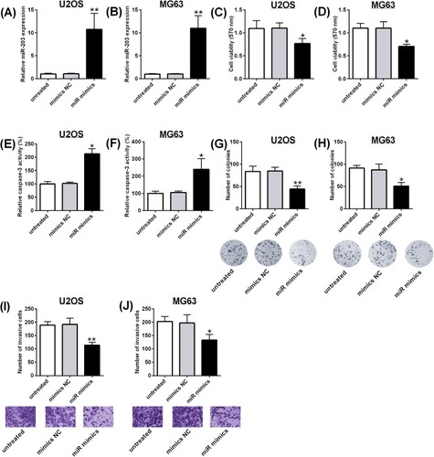
Figure 3 MiR-203 knockdown attenuates the effects of sevoflurane treatment on cell viability, caspase-3 activity and cell invasion of osteosarcoma cells. U2OS and MG63 cells were transfected with inhibitors NC, miR inhibitors or untreated, and 24 hrs later, (A, B) qRT-PCR determined the expression of miR-203 in U2OS and MG63 cells. For in vitro functional assays, U2OS and MG63 cells were exposed to 5% sevoflurane for 6 hrs, and were then transfected with inhibitors NC or miR inhibitors for 24 hrs, (C, D) MTT assay determined the cell viability of U2OS and MG63 cells; (E, F) caspase-3 activity assay kit determined the caspase-3 activity of U2OS and MG63 cells; (G, H) colony formation assay determined the cell growth of U2OS and MG63 cells; (I, J) Transwell invasion assay determined cell invasive ability of U2OS and MG63 cells. N = 3. *P<0.05, **P<0.01 and ***P<0.001.
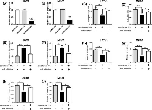
Figure 4 MiR-203 targets 3ʹUTR of WNT2B and inversely regulates WNT2B expression in U2OS cells. (A) Binding sites between miR-203 and 3ʹUTR of WNT2B as predicted by TargetScan were shown. wt = wild type; mut = mutated. (B, C) Luciferase activity of reporter vectors containing WNT2B 3ʹUTR (wt) and WNT2B 3ʹUTR (mut) was determined in U2OS cells with miRNAs transfection. (D) qRT-PCR and Western blot assays determined the WNT2B expression in U2OS cells with miRNAs transfection. (E) U2OS cells were exposed to 5% sevoflurane for 6 hrs and were then transfected with inhibitors NC or miR inhibitors for 24 hrs, and qRT-PCR and Western blot assay determined the WNT2B expression. N = 3. *P<0.05, **P<0.01 and ***P<0.001.
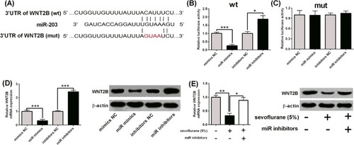
Figure 5 WNT2B overexpression attenuates the effects of sevoflurane treatment on cell viability, caspase-3 activity and cell invasion of osteosarcoma cells. U2OS cells were transfected with pcDNA or pcDNA-WNT2B, and 24 hrs later, (A, B) qRT-PCR and Western blot assays determined the expression of WNT2B in U2OS and MG63 cells. For in vitro functional assays, U2OS cells were exposed to 5% sevoflurane for 6 hrs and were then transfected with pcDNA or pcDNA-WNT2B for 24 hrs, (C) MTT assay determined the cell viability of U2OS cells; (D) caspase-3 activity assay kit determined the caspase-3 activity of U2OS cells; (E) colony formation assay determined the cell growth of U2OS cells; (F) Transwell invasion assay determined cell invasive ability of U2OS cells. N = 3. *P<0.05 and **P<0.01.
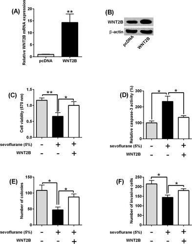
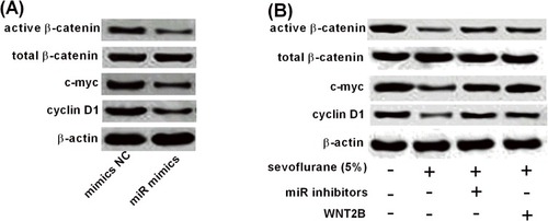
Figure 6 Sevoflurane regulates Wnt/β-catenin signalling via miR-203/WNT2B axis. (A) U2OS cells were transfected with mimic NC or miR mimics, and 24 hrs later, Western blot assay determined protein expression of active β-catenin, total β-catenin, cyclin D1 and c-myc. (B) U2OS cells were exposed to 5% sevoflurane for 6 hrs were then transfected with miRNAs or plasmids for 24 hrs, and Western blot assay determined protein expression of active β-catenin, total β-catenin, cyclin D1 and c-myc.
Data Availability
The data used to support the findings of this study are available from the corresponding author upon request.