Figures & data
Figure 1 Immunohistochemical analysis of STIP1 expression in lung cancer and paracancerous tissue (×400). (A) STIP1 staining was almost undetectable in tissue adjacent to LSCC. (B) Weak STIP1 staining in LSCC tissue. (C) Moderate STIP1 staining in LSCC tissue. (D) Strong STIP1 staining in LSCC tissue. (E) STIP1 staining was almost undetectable in tissue adjacent to LAC. (F) Weak STIP1 staining in LAC tissue. (G) Moderate STIP1 staining in LAC tissue. (H) Strong STIP1 staining in LAC tissue.
Abbreviations: LSCC, lung squamous cell carcinoma; LAC, lung adenocarcinoma.
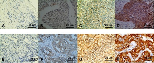
Table 1 Comparison of Average Immunohistochemical Scores Between Lung Squamous Cell Carcinoma and Lung Adenocarcinoma (Score, Mean ± Standard Deviation)
Table 2 Correlation Between STIP1 Expression and Clinicopathological Features in Lung Cancer
Figure 2 STIP1 was strongly expressed at both the mRNA and protein level in lung adenocarcinoma tissue and cells. (A) The STIP1 mRNA expression level in lung adenocarcinoma tissue was significantly higher than that in adjacent tissue (*P<0.05). (B) The STIP1 protein expression level in lung adenocarcinoma tissue was markedly higher than that in adjacent tissue (*P< 0.05). (C) The STIP1 expression level was relatively high in A549 cells. (D) The STIP1 mRNA expression level in A549 cells was considerably higher than that in normal human bronchial epithelial (HBE) cells (*P<0.05). (E) The STIP1 protein expression level in A549 cells was markedly higher than that in HBE cells (*P<0.05).
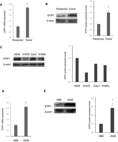
Figure 3 Transfection of STIP1 shRNA enhanced the proliferative, adhesive, and migratory ability, and suppressed the apoptosis, of A549 lung adenocarcinoma cells. (A) Compared with the control and other shRNAs, STIP1 shRNA1 was the most effective at reducing STIP1 expression(*P< 0.05). (B) Transfection of STIP1 shRNA led to a significant decrease in the proliferative ability of lung adenocarcinoma cells(*P< 0.05). (C) Transfection of STIP1 shRNA led to a significant reduction in the adhesive ability of lung adenocarcinoma cells(*P < 0.05). (D) Transfection of STIP1 shRNA led to a marked decline in mobility of lung adenocarcinoma cells(*P < 0.05). (E) Transfection of STIP1 shRNA led to a significant reduction in migratory ability of lung adenocarcinoma cells(*P < 0.05). (F, G) Transfection of STIP1 shRNA led to a significant increase in the apoptosis of lung adenocarcinoma cells(*P < 0.05).
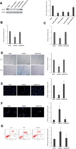
Figure 4 Transfection of STIP1 shRNA led to a reduction in the level of JAK2 and STAT3 phosphorylation and epithelial-to-mesenchymal transition. (A) STIP1 shRNA transfection resulted in reduced levels of p-JAK2 and p-STAT3 (*P< 0.05), but did not affect JAK2 or STAT3 protein levels. (B) Transfection of STIP1 shRNA led to upregulation of E-cadherin expression and suppression of vimentin expression (*P< 0.05).
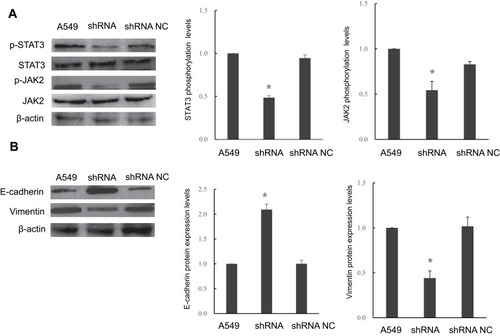
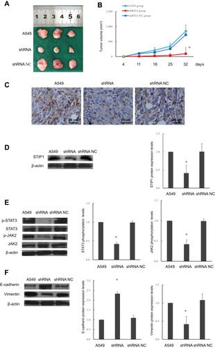
Figure 5 Effects of STIP1 on tumorigenesis in nude mice and associated mechanisms. (A, B) Tumor growth in nude mice injected with A549 lung adenocarcinoma cells transfected with STIP1 shRNA was reduced compared with that in nontransfected cells and those transfected with the negative control (*P< 0.05). (C) Immunohistochemistry indicated that transfection with STIP1 shRNA led to a reduction in STIP1 protein levels in mouse tumors (×400). (D) Western blot showing that STIP1 shRNA suppressed STIP1 protein levels in mouse tumors (*P< 0.05). (E) Transfection with STIP1 shRNA led to a reduction in the levels of phosphorylated JAK2 and STAT3 in STIP1 shRNA-induced tumors in nude mice (*P< 0.05). (F) Transfection with STIP1 shRNA led to the upregulation of E-cadherin expression and suppression of vimentin expression (*P< 0.05).