Figures & data
Figure 1 Detection of proliferation and apoptosis of human esophageal squamous cell carcinoma cells and normal human esophageal epithelial cells. (A) The expression of PRDX1 in esophageal squamous cell carcinoma cells was detected through WB. (B) The activity of the PI3K/AKT pathway in esophageal squamous cell carcinoma cells was detected through WB. (C) Cell proliferation was detected using the MTT method. (D) The plate cloning test was used to test the cloning ability of cells. (E) The rate of apoptosis was detected via flow cytometry. &p <0.05, compared with the HEEC group.
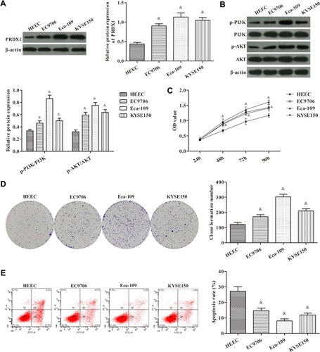
Figure 2 Silencing of PRDX1 expression inhibits the proliferation of Eca-109 cells and promotes apoptosis. (A) The transfection efficiency of PRDX1 siRNA was detected through qRT-PCR. (B) MTT assay for cell proliferation. (C) Plate clone assay for the formation of clones. (D) Flow cytometry for apoptosis. *p<0.05, compared with the control group; #p<0.05, compared with the lv-NC group.
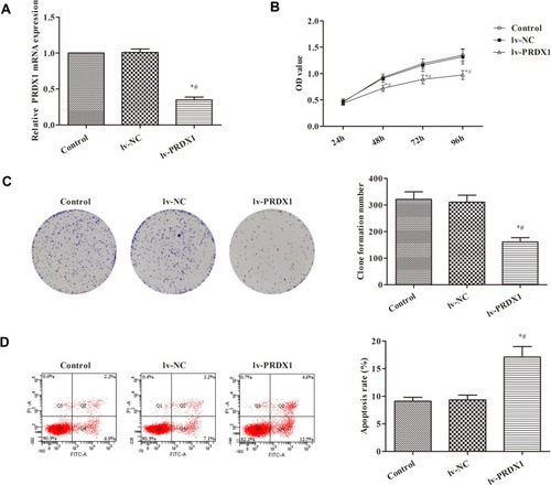
Figure 3 WB assay for the activity of the PI3K/AKT pathway in esophageal squamous cell carcinoma cells. *p<0.05, compared with the control group; #p<0.05, compared with the lv-NC group.
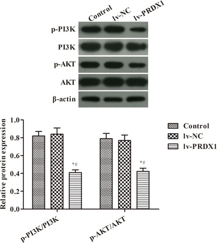
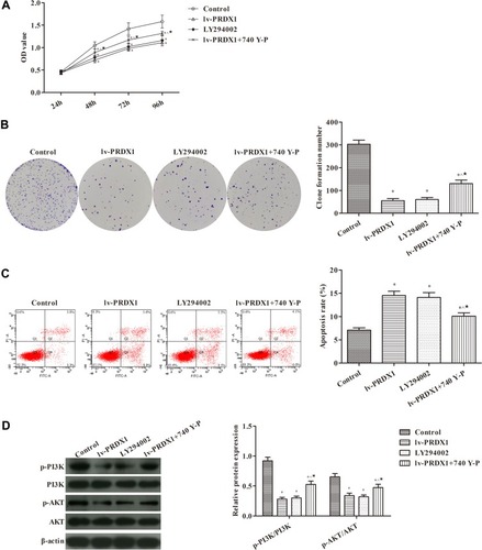
Figure 4 Silencing of PRDX1 expression inhibits the proliferation of Eca-109 cells and promotes apoptosis by decreasing the activity of the PI3K/AKT pathway (A) MTT assay for cell proliferation. (B) Plate clone assay for the formation of clones. (C) Flow cytometry for apoptosis. (D) The activity of the PI3K/AKT pathway in esophageal squamous cell carcinoma was detected through WB. *p<0.05, compared with the control group; ^p<0.05, compared with the lv-PRDX1 group; ★p<0.05, compared with the LY294002 group.