Figures & data
Table 1 Characteristics of the Enrolled Study Population
Table 2 Sensitivity and Accuracy of CEUS, MRI and CT to Diagnose the PCNs
Figure 1 Diagnosis of pancreatic cystic neoplasms by pathology and different modalities.
Abbreviations: PCL, pancreatic cystic lesion; PCN, pancreatic cystic neoplasm; CEUS, contrast-enhanced ultrasound; CT, computed tomography; MRI, magnetic resonance imaging; SCA, serous cystadenoma; MCA, mucinous cystadenoma; IPMN, intraductal papillary mucinous neoplasm; SPN, solid pseudopapillary neoplasm; NEN, neuroendocrine neoplasm; Ca, cystadenocarcinomas.
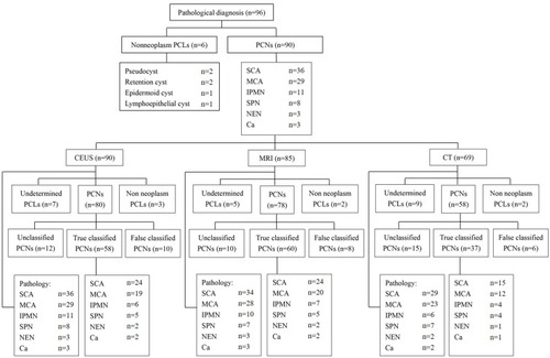
Table 3 Comparison of Diagnostic Accuracy of Different Cysts Size and Detection Rates of Nodule, Septa, Duct Dilatation in Pancreatic Cystic Neoplasms by CEUS, MRI and CT
Table 4 Characteristics of Serous Cystadenomas and Mucinous Cystadenomas Observed by Contrast-Enhanced Ultrasonography
Figure 2 A serous cystadenoma diagnosed via pathology in a 42-year-old woman without symptoms. (A) A US image shows an anechoic-hypoechoic lesion with an irregular shape 3.8 cm in diameter in the pancreatic tail. (B) CEUS image clearly depicts the lobulated shape with a thin septum (red arrow) and a small daughter cyst (yellow arrow). (C) Plain CT scan revealed a watery density mass with an unclear internal structure. (D) Contrast-enhanced CT demonstrates lobulated morphology and septum (red arrow). (E) T2-weighted MRI shows a thin intralesional septum (red arrow). (F) Enhanced MRI reveals the septum (red arrow).
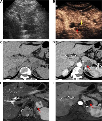
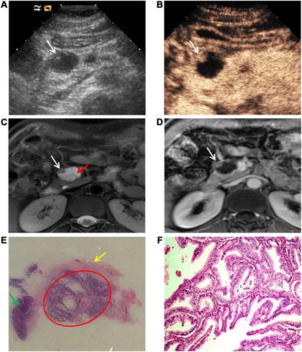
Figure 3 A mucinous cystadenocarcinoma diagnosed via pathology in a 48-year-old woman without symptoms. (A) A US image shows a cystic lesion with a hypoechoic attachment (white arrow) 2.5 cm in diameter in the pancreatic head. (B) A CEUS image clearly displays the round margin and a nodule in the cyst (white arrow) with enhancement. (C) T2-weighted MRI confirms presence of lesion and shows a nodule (white arrow) and thin intralesional septa (red arrow). (D) Enhanced MRI reveals the nodule (white arrow). (E) Photograph of pathologic specimen through HE staining reveals a nodule (red circle), cyst wall (yellow arrow), and pancreas parenchyma (green arrow). (F) Malignant tumor cells as shown under high power microscope.