Figures & data
Table 1 Clinical and Histopathologic Data of Patients in the modelling group (Mean±Standard Deviation)
Table 2 Clinical and Histopathologic Data of Patients in the validation group (Mean±Standard Deviation)
Figure 1 A 50-year-old man had a peripheral pulmonary lesion in the right lung. (A) A round lesion (arrow) could be seen in the right middle lobe with a size of 9 mm×9 mm. (B) Endobronchial ultrasonography revealed a heterogeneous lesion with hyper- and hypoechoic areas. (C) Tumor and normal cells existed in the same area with necrosis and fibrosis in the middle (HE ×100).
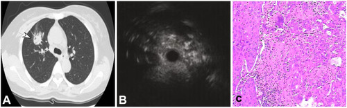
Figure 2 A 65-year-old woman had a peripheral pulmonary lesion (inflammatory pseudotumor) in the lung back segment. (A) A lesion of 30 mm×28 mm (arrow) could be seen in the back segment with an air bronchogram. (B) Endobronchial ultrasound could detect a heterogeneous lesion with diffuse short signals. (C) A few distorted and expanded bronchia existed in the inflammatory cells and normal cells (HE ×100).
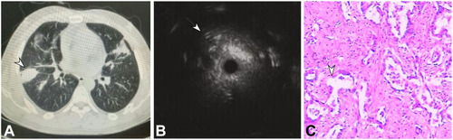
Table 3 Comparisons of Clinical Factors and EBUS Image Patterns in Predicting Malignancy in Patients
Table 4 Logistic Model for Predicting Peripheral Lung Cancer Using EBUS and Clinical Features
Table 5 Testing of the Logistic Equation with Pathological Results in 50 Patients
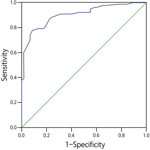
Figure 3 Receiver operating characteristic curve for the direction model for diagnosis of malignant peripheral pulmonary lesions (PPLs). The prediction model was developed based on data from 135 consecutive patients.