Figures & data
Figure 1 PSMA3-AS1 is upregulated in lung cancer. (A) PSMA3-AS1 expression was increased in lung cancer tissues compared to adjacent normal tissues. (B) PSMA3-AS1 expression was higher in lung cancer tissues with advanced stages. (C) PSMA3-AS1 level was higher in lung cancer tissues with metastasis. (D) Relative expression of PSMA3-AS1 in lung cancer cell lines. (E) PSMA3-AS1 high expression correlated with a low survival rate. Low expression group: 18 samples. High expression group: 23 samples. *P<0.05.
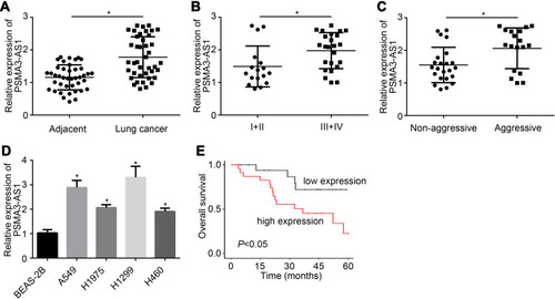
Figure 2 PSMA3-AS1 knockdown suppresses proliferation, migration and invasion. (A) PSMA3-AS1 expression was suppressed after transfection of si-PSMA3-AS1. (B and C) CCK8 assay was performed to analyze proliferation. (D) PSMA3-AS1 knockdown led to decreased colony numbers. (E and F) PSMA3-AS1 knockdown inhibited cell migration and invasion. (G) CCK8 assay for cell proliferation. (H and I) Transwell assay for cell migration and invasion after PSMA3-AS1 overexpression. *P<0.05.
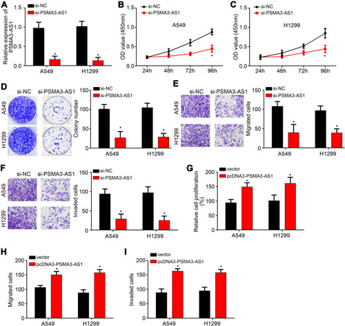
Figure 3 PSMA3-AS1 inhibits miR-4504 in lung cancer. (A) Diagram for the binding between PSMA3-AS1 and miR-4504. (B and C) Luciferase reporter assay showed that miR-4504 inhibited the luciferase activity of WT PSMA3-AS1. (D) RNA pulldown assay indicated that PSMA3-AS1 was enriched by biotin-labeled miR-4504-WT. (E) PSMA3-AS1 overexpression suppressed the level of miR-4504. (F) MiR-4504 was downregulated in lung cancer tissues. (G) PSMA3-AS1 expression was negatively correlated with miR-4504 in lung cancer tissues. *P<0.05.
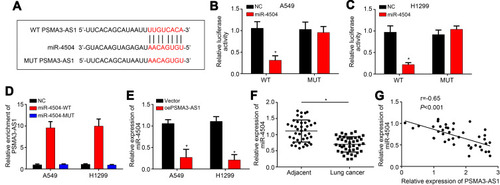
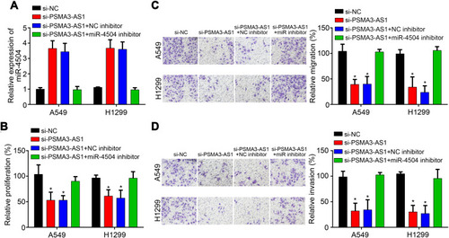
Figure 4 PSMA3-AS1 promotes lung cancer progression via inhibiting miR-4504. (A) Relative expression of miR-4504 was determined via qRT-PCR. (B) Proliferation was analyzed via CCK8 assay. (C and D) Transwell assay was conducted to determine migration and invasion after transfection of indicated vectors. *P<0.05.