Figures & data
Table 1 The Clinicopathological Features of These Patients (n = 30)
Table 2 Primers for PCR
Figure 1 CircGDI2 expression was decreased in OSCC tissues and cells. (A and B) CircGDI2 expression by qRT-PCR in 30 pairs of OSCC tissues and matched normal tissues, HOK, CAL-27 and SCC-15 cells. Blots were representative of n = 3. (C and D) The levels of circGDI2 and GDI2 linear mRNA by qRT-PCR in RNase R-treated RNA extracts from CAL-27 and SCC-15 cells. Blots were representative of n = 6. (E and F) The subcellular localization of circGDI2 in both CAL-27 and SCC-15 cells. Error bars indicated SD of triplicate experiments. (G) The levels of lamin B and α-tubulin by Western blot in cytoplasmic and nuclear fractionations of CAL-27 and SCC-15 cells. A representative experiment was shown in triplicate. *P < 0.05.
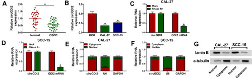
Figure 2 CircGDI2 was transferred by exosomes in OSCC cells. (A) The representative micrograph of the exosomes derived from CAL-27 and SCC-15 cells by TEM (scale bars=100 nm). Red arrows pointed the exosomes. (B and C) The levels of CD63 and CD9 by Western blot in the exosomes and cell lysates. GAPDH was used as a negative control. A representative experiment was shown in triplicate. (D) CAL-27 cells were treated with 30 µg/mL of the exosomes derived from CAL-27 cells transfected with pcDNA-NC, pcDNA-circGDI2, si-NC or si-circGDI2, and then circGDI2 expression was assessed by qRT-PCR in the Donor cells, the exosomes and Recipient cells. Blots were representative of n = 6. (E) SCC-15 cells were treated with 30 µg/mL of the exosomes derived from SCC-15 cells transfected with pcDNA-NC, pcDNA-circGDI2, si-NC or si-circGDI2, followed by the determination of circGDI2 level in the Donor cells, the exosomes and Recipient cells. Blots were representative of n = 6. *P < 0.05.
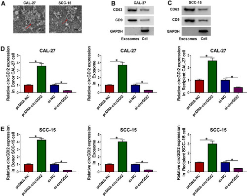
Figure 3 The elevated expression of exosomal circGDI2 hampered OSCC cell proliferation, migration, invasion and glycolysis. CAL-27 and SCC-15 cells (Recipient cells) were treated with 30 µg/mL of the exosomes from pcDNA-NC- or pcDNA-circGDI2-transfected Donor cells. (A and B) MTT assay for cell proliferation. (C and D) Transwell assay for cell migration and invasion. (E–H) A corresponding assay kit for glucose consumption and lactate production. (I and J) Measurement of ECAR in treated Recipient cells. (K and L) Western blot for GLUT1 and LDHA levels in treated Recipient cells. Error bars indicated SD of triplicate experiments. *P < 0.05.
Abbreviations: pcDNA-NC-exo, exosomes derived from OSCC cells transfected with pcDNA; pcDNA-circGDI2-exo, exosomes derived from OSCC cells transfected with pcDNA-circGDI2.
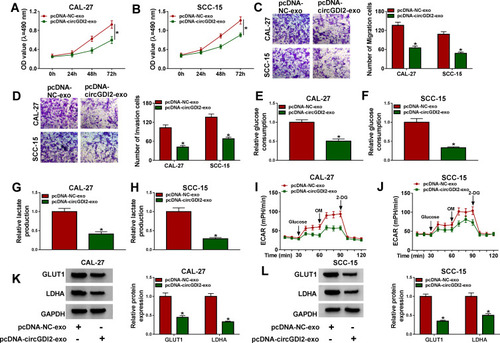
Figure 4 CircGDI2 acted as a sponge of miR-424-5p. (A) Schematic model of the miR-424-5p-binding sequence within circGDI2 and the mutant of the binding sequence. (B and C) The luciferase activity in CAL-27 and SCC-15 cells cotransfected with circGDI2 luciferase reporter vector (circGDI2 WT) or the site-directed mutant (circGDI2 MUT) and miR-424-5p mimic or miR-NC mimic. Blots were representative of n = 6. The expression of miR-424-5p by qRT-PCR in CAL-27 and SCC-15 cells treated with pcDNA-NC-exo or pcDNA-circGDI2-exo (D and E), in 30 pairs of OSCC tissues and matched normal tissues (F), HOK, CAL-27 and SCC-15 cells (G). Blots were representative of n = 3. (H) Correlation between circGDI2 level and miR-424-5p expression in OSCC tissues using the Spearman test. *P < 0.05.
Abbreviations: pcDNA-NC-exo, exosomes derived from OSCC cells transfected with pcDNA; pcDNA-circGDI2-exo, exosomes derived from OSCC cells transfected with pcDNA-circGDI2.
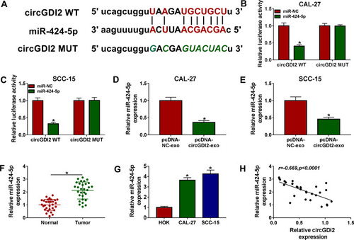
Figure 5 The suppressive impact of exosomal circGDI2 overexpression on OSCC cell malignant behaviors was reversed by the restored level of miR-424-5p. CAL-27 and SCC-15 cells (Recipient cells) were transfected with miR-424-5p mimic or miR-NC mimic and then treated with 30 µg/mL of the exosomes from pcDNA-NC- or pcDNA-circGDI2-transfected Donor cells, followed by the detection of miR-424-5p expression by qRT-PCR (A and B), cell proliferation by MTT assay (C and D), cell migration and invasion by transwell assay (E and F), glucose consumption and lactate production using a corresponding assay kit (G and H), ECAR using a standard assay (I and J), LDHA and GLUT1 levels by Western blot (K and L). A representative experiment was shown in triplicate. *P < 0.05.
Abbreviations: pcDNA-NC-exo, exosomes derived from OSCC cells transfected with pcDNA; pcDNA-circGDI2-exo, exosomes derived from OSCC cells transfected with pcDNA-circGDI2.
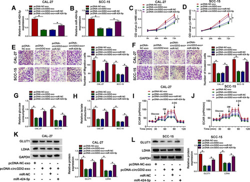
Figure 6 SCAI was a direct target of miR-424-5p. (A) Schematic of the miR-424-5p-binding sites within SCAI 3ʹUTR identified by the starBase v.2 software and mutated miR-424-5p-binding sites. (B and C) The luciferase activity in CAL-27 and SCC-15 cells cotransfected with SCAI 3ʹUTR WT or SCAI 3ʹUTR MUT and miR-424-5p mimic or miR-NC mimic. Blots were representative of n = 6. SCAI mRNA expression by qRT-PCR and protein level by Western blot in CAL-27 and SCC-15 cells transfected with anti-miR-NC or anti-miR-424-5p (D–G), OSCC tissues and matched normal tissues (H and I), HOK, CAL-27 and SCC-15 cells (J and K). Blots were representative of n = 3. (L) Correlation between SCAI mRNA level and miR-424-5p expression in OSCC tissues using the Spearman test. *P < 0.05.
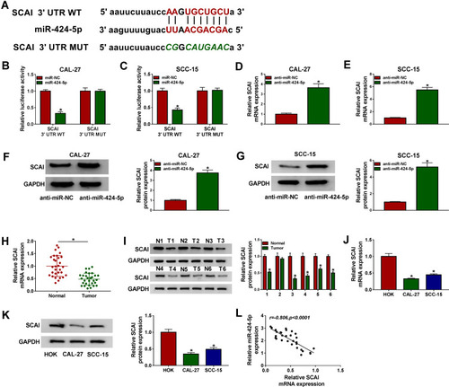
Figure 7 The mitigative effects of miR-424-5p knockdown on OSCC cell malignant behaviors were mediated by SCAI. CAL-27 and SCC-15 cells were transfected with anti-miR-NC, anti-miR-424-5p, anti-miR-424-5p+si-NC or anti-miR-424-5p+si-SCAI. (A) SCAI expression by Western blot in transfected cells. (B and C) Cell proliferation by MTT assay. (D and E) Cell migration and invasion by transwell assay. (F and G) Glucose consumption and lactate production using a corresponding assay kit. (H and I) ECAR using a standard assay. (J and K) LDHA and GLUT1 levels by Western blot in transfected cells. A representative experiment was shown in triplicate. *P < 0.05.
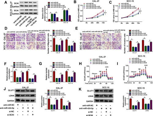
Figure 8 Exosomal circGDI2 regulated SCAI expression through sponging miR-424-5p. (A) Correlation between circGDI2 expression and SCAI mRNA level in OSCC tissues using the Spearman test. CAL-27 and SCC-15 cells (Recipient cells) were transfected with miR-424-5p mimic or miR-NC mimic and then treated with 30 µg/mL of the exosomes from pcDNA-NC- or pcDNA-circGDI2-transfected Donor cells, followed by the measurement of SCAI mRNA expression by qRT-PCR (B and C), SCAI protein level by Western blot (D and E). Blots were representative of n = 6. *P < 0.05.
Abbreviations: pcDNA-NC-exo, exosomes derived from OSCC cells transfected with pcDNA; pcDNA-circGDI2-exo, exosomes derived from OSCC cells transfected with pcDNA-circGDI2.
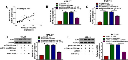
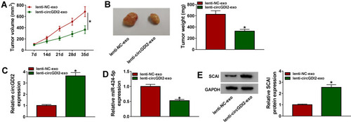
Figure 9 Overexpression of exosomal circGDI2 hampered tumor growth in vivo. CAL-27 cells were subcutaneously injected into the nude mice (n= 6 each group). One week later, intratumor injection for the exosomes from lenti-NC- or lenti-circGDI2-transduced CAL-27 cells was performed. After 35 days cell implantation, all mice were euthanized. (A) Tumor volume measurement began on 7 days after implantation and was conducted every week. (B) Representative pictures and average weight of the xenograft tumors. (C–E) The levels of circGDI2, miR-424-5p and SCAI were assessed by qRT-PCR or Western blot in excised tumor tissues. A representative experiment was shown in triplicate. *P < 0.05.