Figures & data
Figure 1 Sevoflurane regulated the abilities of viability, apoptosis, migration, invasion, and suppressed the β-catenin signaling in glioma cells. (A and B) MTT assay was used to manifest the cell viability of U251 or U343 cells at 24 h, 48 h, and 72 h after treatment of sevoflurane for various concentrations (1%, 2%, 4%). (C) Flow cytometry was carried out to measure the rate of apoptosis when cells administrated with sevoflurane. (D and E) Transwell assay was applied to evaluate migration and invasion abilities of U251 and U343 cells after treatment with sevoflurane (magnification: 100x, n=3). (F and G) Western blot assay was used to detect the expression of β-catenin signaling-related proteins, namely β-catenin, c-Myc, CyclinD1 in glioma cells. *P<0.05.
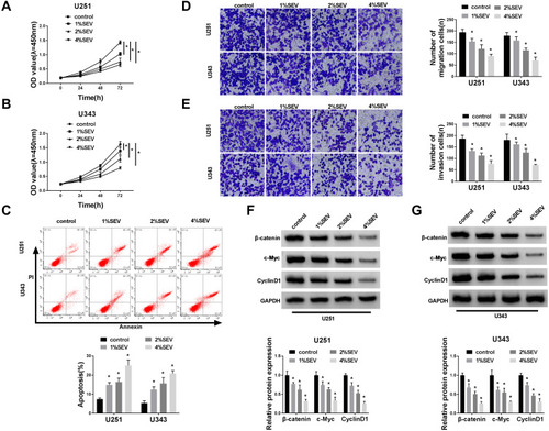
Figure 2 Overexpression of miR-218-5p repressed cell viability, migration, invasion, and β-catenin signaling, but promoted the apoptosis in glioma cells in vitro. (A) The mRNA expression level of miR-218-5p of U251 and U343 cells after treatment with sevoflurane was detected by qRT-PCR assay. U251 and U343 cells were transfected with miR-NC mimics or miR-218-5p mimics. (B) The miR-218-5p expression was tested via qRT-PCR assay at 48 h post-transfection in U251 and U343 cells. (C–E) The cell viability of U251 and U343 cells was measured by MTT assay at 24 h, 48 h and 72 h after transfection (C and D) and the rate of apoptosis was explored by flow cytometry after transfection for 48 h as well (E). (F and G) The number of migration and invasion cells was counted at 48 h following transfection through transwell chamber experiment (magnification: 100x, n=3). (H and I) The gray value (H) and analysis results (I) of protein immunoblots of β-catenin signal pathway-related proteins were shown.*P<0.05.
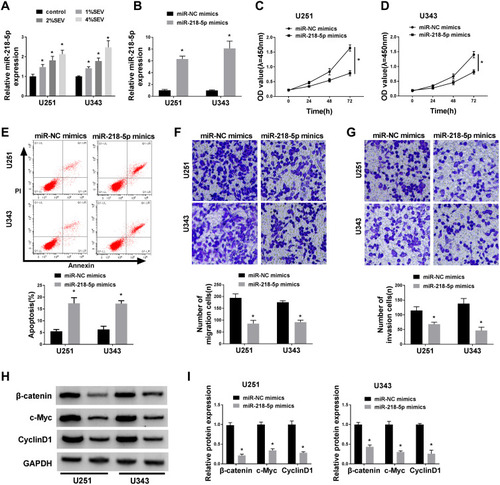
Figure 3 Sevoflurane regulated the cell viability, apoptosis, migration, invasion, and activation of β-catenin signaling by targeting miR-218-5p. (A) qRT-PCR assay showed the expression level of miR-218-5p in U251 and U343 cells after treatment with sevoflurane and miR-NC inhibitors or miR-218-5p inhibitors. (B and C) MTT assay showed cell viability of U251 and U343 cells after treatment. (D) Flow cytometry assay witnessed cell apoptosis of U251 and U343 cells after administration. (E and F) Transwell assay exhibited the number of migration and invasion cells after treatment (magnification: 100x, n=3). (G and H) Western blot analysis measured the activation of β-catenin signal pathways. *P<0.05.
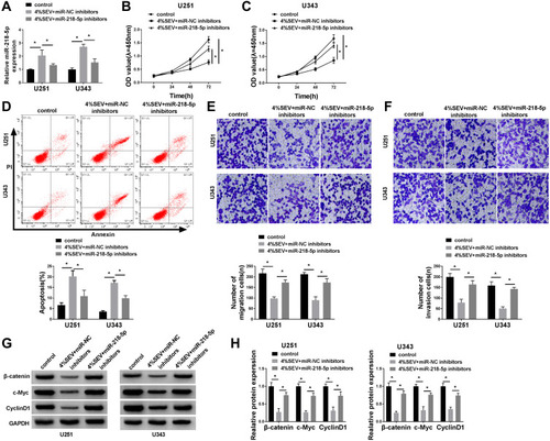
Figure 4 DEK was a direct target of miR-218-5p and negatively regulated by miR-218-5p. (A) The putative binding site and the mutant sites in of miR-218-5p on the 3ʹ-UTR of DEK mRNA were shown. (B–E) U251 and U343 cells were transfected with DEK-WT or DEK-MUT and miR-218-5p mimics or miR-NC mimics, miR-218-5p inhibitors or miR-NC inhibitors, followed by the measure of dual-luciferase activities at 48 h after transfection. (F–I) The protein level of DEK in the two cell lines was tested by Western blot after transfection. *P<0.05.
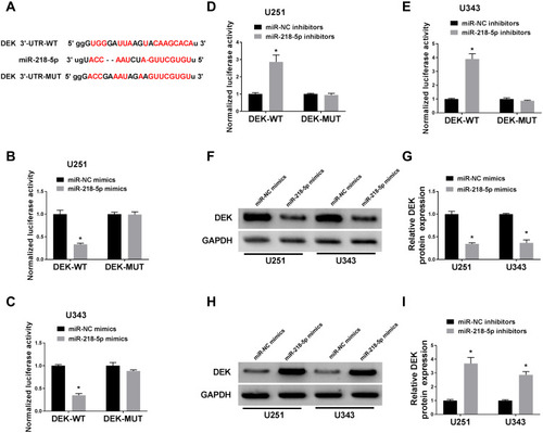
Figure 5 MiR-218-5p restored the cell viability, apoptosis, migration, invasion abilities and revitalized β-catenin signaling by targeting DEK. (A and B) Western blot assay revealed the protein level of DEK in U251 and U343 cells after co-transfection. (C and D) MTT assay showed cell viability of U251 and U343 cells after co-transfection. (E) Flow cytometry assessment performed cell apoptosis of U251 and U343 cells after treatment. (F and G) Transwell assay exhibited migration and invasion abilities of the two cell lines (magnification: 100x, n=3). (H and I) Western blot analysis uncovered the expression patterns upon β-catenin, c-Myc, CyclinD1 in glioma cells. *P<0.05.
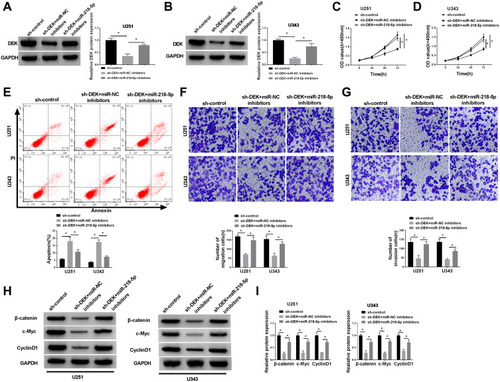
Figure 6 Upregulation of DEK inverted sevoflurane-mediated repression on cell viability, migration, and invasion and β-catenin signal pathway, but promotion on apoptosis in glioma cells. U251 and U343 cells were transfected with pcDNA3.1 vector or pcDNA3.1-DEK after treatment with 4% sevoflurane. (A and B) The protein level of DEK in U251 and U343 cells, (C and D) cell viability of U251 and U343 cells and apoptosis (E), migration (F), and invasion (G) of cell numbers in U251 and U343 cells, supplemented with the expression of β-catenin signal pathway-associated proteins (H and I) were shown.*P<0.05.
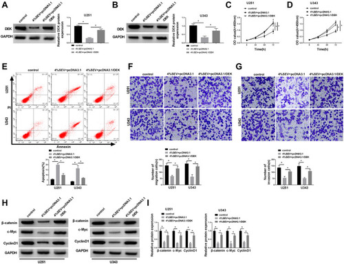
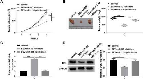
Figure 7 Knockdown of miR-218-5p reversed sevoflurane-treated effects on tumor growth in vivo. U251 cells transfected with in-miR-NC or in-miR-218-5p were injected into the nude mice for 3 days, mice were injected of 50 mg/kg sevoflurane or 0.5 mL soybean oil for 25 days. After the mice were sacrificed, the tumor was measured. Tumor volume (A) and Tumor weight (B) were detected. (C) The expression level of miR-218-5p was detected by qRT-PCR. (D) The protein expression level of DEK was measured by Western blot assay. ***P<0.001.