Figures & data
Figure 1 Flowchart of patients selection process.
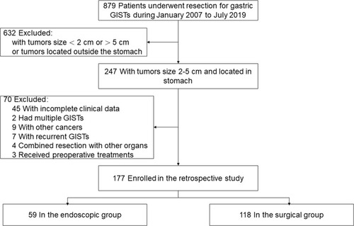
Figure 2 Laparoscopic resection of gastric GIST. (A) Direct resection by liner stapler. (B) Open the stomach cavity and cut with ultrasonic scalpel along the edge of the tumor, when the bottom of the tumor cannot be identified from outside of the stomach. (C) Postoperative specimen and the results of the final postoperative examination show this tumor is GIST with the size< 5cm.
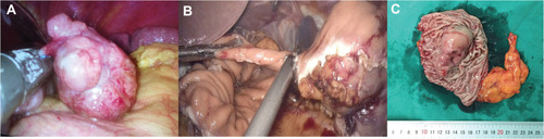
Figure 3 A 65 years old woman, complained upper abdominal distention for 3 months. (A) A submucous lesion (about 3.5x2.0 cm in size) was found in the body of stomach by gastroscopy. (B) She received an EFTR (endoscopic full-thickness resection) procedure under an experienced endoscopist. (C) The wound in stomach was large and deep after the lesion was removed. (D and E) The wound was closed by detachable snare loop technique. (F) The specimen of the removed lesion, with the size about 3.0x4.5 cm, and the GIST diagnosis was confirmed by the pathologist.
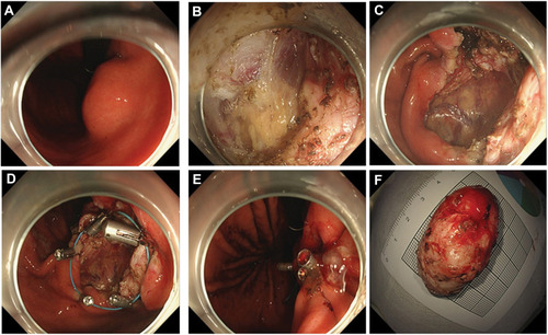
Table 1 Demographic and Clinical Characteristics of Two Groups
Table 2 Pathological Characteristics of Two Groups
Table 3 Genetic Characteristics of Two Groups
Table 4 Short-Term Outcomes of Two Groups
Table 5 Data of the Complicated Cases in Two Groups
Table 6 Causes of Reoperation of Two Groups
Table 7 The Complications and Reoperation of 2–3cm Gastric GISTs in Two Groups
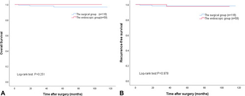
Figure 4 The difference between two groups of patients in overall survival (A) or recurrence-free survival (B) did not attain statistical significance (P=0.251 and P=0.978, respectively).