Figures & data
Figure 1 Case screening flow chart.
Notes: A total of 576 GISTs cases in our center (Xiangya Hospital) from May 2017 to Oct 2019 were screened. One hundred and eighty-five patients with no available genetic data of routine Sanger sequencing and 11 patients lost to follow-up were all excluded. Twenty-nine patients with poor quality FFPE were also excluded. Finally, 23 cases of 52 wild-types KIT/PDGFRA GISTs were included (negative for exons 9,11,13,14, and 17 in KIT and exon 12, 14, and 18 in PDGFRA using Sanger sequencing). A total of 136 GISTs cases between May 2017 to May 2020 in the Gene+ database and the 131 cases of GISTs from the MSKCC database were also included in our study.
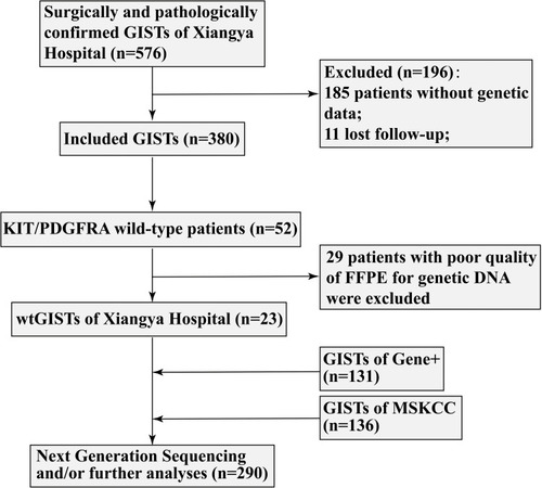
Table 1 Clinical Characteristics of GIST with NF1 Mutation
Table 2 The Mutation Types and Their Differences Between mNF1 with mKIT and mNF1 with wtKIT
Table 3 Concurrent Mutation of NF1 and KIT in GISTs Patients
Figure 2 The mutation loci and subtypes within the NF1 gene and their corresponding pathogenicity in GISTs.
Notes:#Xiangya Hospital; ##Gene+ database; ###MSKCC database; ±, Neutral; –, Deleterious; Red symbols of #, ## and ###represent n=2.
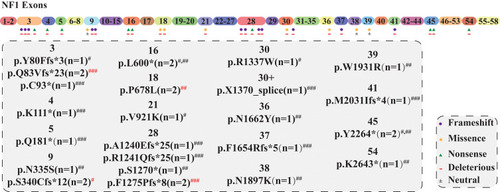
Table 4 NF1 Concurrent with KIT Mutation
Figure 3 KIT mutations loci and subtype.
Notes: The distribution of various mutation subtypes identified by NGS within the exons of KIT gene; #Xiangya Hospital; ##Gene+ database; ###MSKCC database; ±, Neutral; –, Deleterious; Red symbols of ## and ###represent n=2.
Abbreviations: TM, transmembrane domain; JM, juxtamembrane domain; N, number.
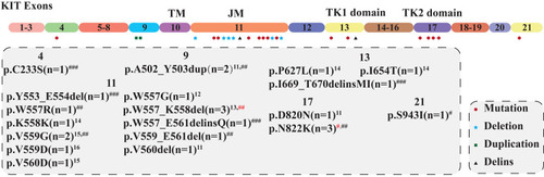
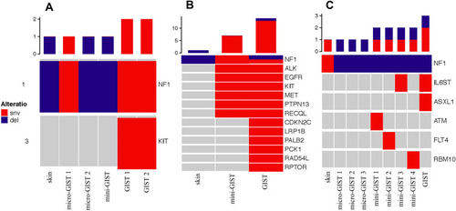
Figure 4 The genomic landscapes of skin neurofibroma and GISTs in 3 cases of type I neurofibromatosis.
Notes: (A–C) represent individual cases 1–3, respectively. In panel A (case 1) and panel B (case 2), compared with the lesions of skin neurofibromas and micro-GISTs (≤1 cm) that merely harbor mNF1, GISTs lesions (>2 cm) have NF1 aberration with a concurrent mKIT. In all three cases, the number of gene mutations is gradually increasing with the neurofibromas lesions to micro-GISTs or/and mini-GISTs to clinic GISTs.