Figures & data
Table 1 Primers Used in qRT-PCR
Figure 1 CircEIF6 is highly expressed in pancreatic cancer tissues and cell lines. (A) The level of circEIF6 in tumor tissues (n=39) and adjacent normal tissues (n=39) from pancreatic cancer patients was evaluated by qRT-PCR. (B) The expression of circEIF6 in the tumor tissues from pancreatic cancer patients in I+II phase and III phase was assessed by qRT-PCR. (C) ROC curve was used to analyze the diagnostic value of circEIF6 in pancreatic cancer patients. (D) qRT-PCR was used to measure the expression of circEIF6 in normal human pancreatic duct epithelial cell line (HPDE) and two pancreatic cancer cell lines (Hs 766T and SW1990). (E and F) The stability of circEIF6 along with its linear form EIF6 mRNA was assessed using RNase R by qRT-PCR. *P<0.05.
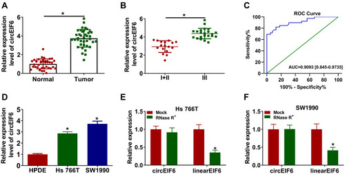
Figure 2 CircEIF6 knockdown hampers cell proliferation, migration, invasion and induces cell apoptosis of pancreatic cancer cells. (A–H) Hs 766T and SW1990 cells were transfected with si-EIF6 or its control si-NC, and untreated pancreatic cancer cells were used as the control group. (A and B) The expression of circEIF6 was evaluated by qRT-PCR. (C and D) Transfected pancreatic cancer cells in 96-well plates were incubated with CCK8 after fixed time interval following transfection to evaluate the number of viable cells, and cell proliferation curve was generated. (E and F) Cell migration ability and cell invasion ability were assessed by transwell assays. The representative images of migrated or invaded cells in different groups are shown. (G) The apoptosis rate (the first quadrant and the fourth quadrant) was analyzed by flow cytometry. (H) The levels of p-AKT, AKT, p-PI3K and PI3K were examined by Western blot assay, and the intensities of protein bands were determined by ImageJ software. *P<0.05.
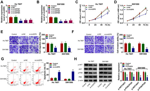
Figure 3 MiR-557 is a target of circEIF6 in pancreatic cancer cells. (A) Circular RNA Interactome was used to predict the targets of circEIF6 based on the complementary sequence between circEIF6 and candidate targets. The diagram revealed the seed sequence of miR-557 which was complementary with the matched sequence in circEIF6. (B and C) Dual-luciferase reporter assay was conducted to confirm the interaction and the target sequence between circEIF6 and miR-557. (D) The level of miR-557 in pancreatic cancer tissues (n=39) along with paired normal tissues (n=39) was evaluated by qRT-PCR. (E) Pearson correlation coefficient was used to assess the linear correlation between the expression of miR-557 and circEIF6. (F) The expression of miR-557 in HPDE, Hs 766T and SW1990 was determined by qRT-PCR. (G) Hs 766T and SW1990 cells were transfected with si-NC or si-circEIF6, and evaluation of miR-557 expression was performed by qRT-PCR. *P<0.05.
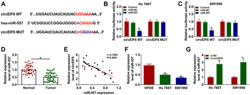
Figure 4 CircEIF6 knockdown restrains the malignant phenotypes of pancreatic cancer cells through enhancing miR-557 level. (A–H) Hs 766T and SW1990 cells were transfected with si-EIF6 alone or together with in-miR-557. (A) Evaluation of miR-557 abundance in transfected pancreatic cancer cells was conducted via qRT-PCR. (B and C) The influences of circEIF6 silencing and miR-557 silencing on the proliferation of pancreatic cancer cells were analyzed by CCK8 assay. (D and E) Transwell assays were implemented to analyze cell migration and invasion abilities in transfected pancreatic cancer cells. (F) Flow cytometry was conducted to count the percentage of apoptotic pancreatic cancer cells (the first quadrant and the fourth quadrant). (G and H) Western blot assay was conducted to detect the abundance of p-AKT, AKT, p-PI3K and PI3K in transfected pancreatic cancer cells. *P<0.05.
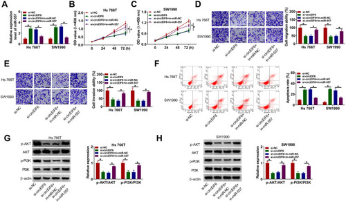
Figure 5 SLC7A11 is a target of miR-557 in pancreatic cancer cells. (A) The interacted mRNAs of miR-557 were predicted through using bioinformatic software (TargetScan), and SLC7A11 was predicted to interact with miR-557 via its “UGCAAA” sequence. (B and C) The target interaction between miR-557 and SLC7A11was verified by dual-luciferase reporter assay. (D and E) qRT-PCR and Western blot assay were conducted to determine the mRNA and protein expression of SLC7A11 in pancreatic tumor tissues and adjacent normal tissues. (F) Evaluation of SLC7A11 level in HPDE, Hs 766T and SW1990 was conducted by Western blot assay. (G and H) The linear relationship between SLC7A11 and miR-557 or circEIF6 was analyzed by Pearson correlation coefficient. (I) Hs 766T and SW1990 cells were transfected with miR-NC or miR-557, and the level of miR-557 was assessed by qRT-PCR after transfection for 24 hours. (J) Western blot assay was used to analyze the protein expression of SLC7A11 in Hs 766T and SW1990 cells transfected with miR-NC or miR-557. (K) Evaluation of SLC7A11 protein expression in pancreatic cancer cells transfected with si-NC, si-circEIF6, si-EIF6 + in-miR-NC or si-EIF6 + in-miR-557 was conducted by Western blot assay. *P<0.05.
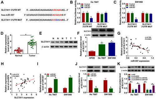
Figure 6 SLC7A11 overexpression partly alleviates miR-557-mediated effects in pancreatic cancer cells. (A-H) Hs 766T and SW1990 cells were transfected with miR-NC, miR-557, miR-557 + pcDNA or miR-557 + SLC7A11. (A) The protein level of SLC7A11 in transfected pancreatic cancer cells was examined by Western blot assay. (B and C) CCK8 assay was utilized to evaluate cell proliferation ability. (D and E) Transwell assays were conducted to assess cell migration and invasion abilities. (F) The percentage of apoptotic cells (the first quadrant and the fourth quadrant) was evaluated by flow cytometry. (G and H) The levels of p-AKT, AKT, p-PI3K, PI3K were determined by Western blot assay. *P<0.05.
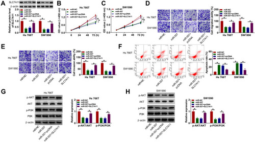
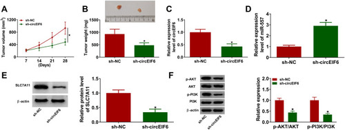
Figure 7 CircEIF6 interference suppresses the growth of pancreatic cancer xenograft tumors in vivo. (A) Tumor volume was calculated every 7 days according to the formula of length×width2×0.5. (B) Tumors were weighed after euthanizing mice at 28-day post-inoculation. (C) The expression of circEIF6 in tumor tissues from SW1990 cells transfected with sh-NC or sh-EIF6 was detected by qRT-PCR. (D) The level of miR-557 was measured by qRT-PCR. (E) Western blot assay was used to analyze the protein level of SLC7A11 in tumor tissues. (F) The levels of p-AKT, AKT, p-PI3K and PI3K were determined by Western blot assay. *P<0.05.