Figures & data
Figure 1 CRC tissues exhibited altered expression of circFAT1 and mature miR-10a. Tissue samples from a total of 64 CRC patients were subjected to total RNA extractions and RT-qPCRs to analyze the expression of circFAT1 (A), mature miR-10a (B) and premature miR-10a (C) in CRC. Heatmaps plotted using Heml 1.0 were used to express differential gene expression in paired tissues.
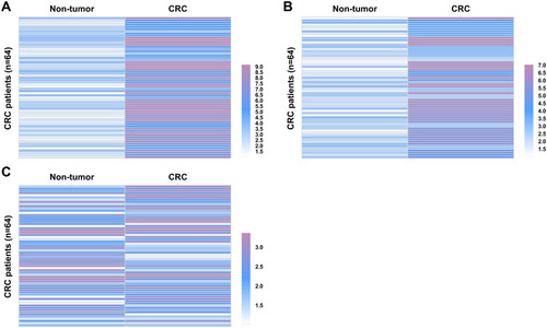
Figure 2 MiR-10a was positively and significantly correlated with circFAT1. The correlations between circFAT1 and mature miR-10a (A) or premature miR-10a (B) across both CRC tissues were analyzed by Pearson’s correlation coefficient.
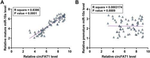
Figure 3 CircFAT1 overexpression decreased miR-10a maturation in both WiDr and HT-29 cells. The close correlation between circFAT1 and mature miR-10a across CRC tissues indicated the possible role of circFAT1 in regulating miR-10a maturation in CRC. To explore this possibility, WiDr and HT-29 cells were transfected with either circFAT1 expression vector or miR-10a mimic, followed by the confirmation of the overexpression of circFAT1 and mature miR-10a every 24h until 144h (A). The effects of circFAT1 overexpression on the expression of mature miR-10a (B) and premature miR-10a (C), and the effects of mature miR-10a overexpression on circFAT1 expression (D) were also analyzed by RT-qPCR. *p<0.05.
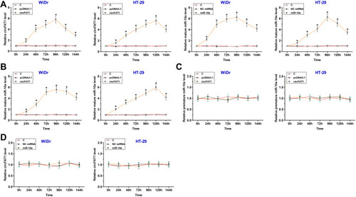
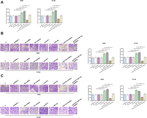
Figure 4 CircFAT1 overexpression of increased CRC cell proliferation, invasion, and migration through miR-10a. The role of circFAT1 and miR-10a in regulating CRC cell proliferation, invasion, and migration were analyzed by BrdU assay (A) and Transwell invasion (B) and migration (C) assays, respectively. *p<0.05.