
Figures & data
Table 1 A Summary of Demographic Data, Clinical Features, Treatments and Outcomes of the Patients
Figure 1 The distribution of symptoms for all patients.
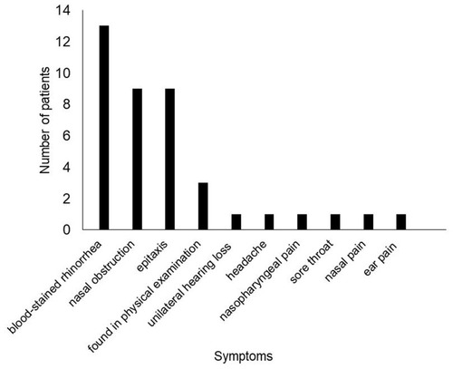
Figure 2 Nasopharyngeal endoscopy revealed a pedicled polypoid mass was located on the root of the nasopharynx.
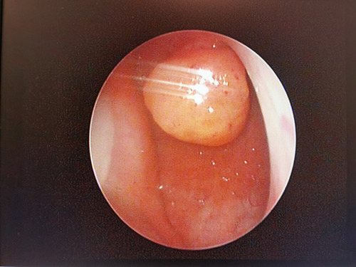
Figure 3 Sinus computer tomography images showed the tumor was pedicled and enhanced at the posterior margin of the nasal septum on the axial images (A, arrow) and the reconstructed coronal images (B, arrow), invading the roof of the nasopharynx slightly. No bone destruction was found (C, arrow). The tumor was demonstrated a moderate signal in T1-weighted magnetic resonance images (D, arrow), and a slightly high signal in T2-weighted magnetic resonance images (E, arrow). It was strongly enhanced in enhanced images (F–H, arrow).
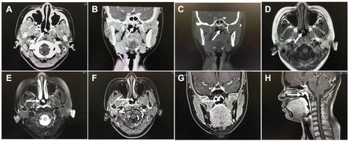
Figure 4 The tumor displayed papillary structures with a hyalinized fibrovascular cores and a prominent intervening spindle cell component ((A), H&E staining, x100). The papillae were lined by bland columnar epithelium with elongated slightly crowded nuclei which displayed fine chromatin and absent to inconspicuous nucleoli ((B), H&E staining, x200). Immunophenotype for TL-LGNPPA showed both tumor cells were strongly positive for CK7 and TTF ((C and D), immunohistochemical [IHC], x200), and negative for TG ((E), immunohistochemical [IHC], x200). The Ki-67 proliferation value was about 1% ((F), immunohistochemical [IHC], x200).
![Figure 4 The tumor displayed papillary structures with a hyalinized fibrovascular cores and a prominent intervening spindle cell component ((A), H&E staining, x100). The papillae were lined by bland columnar epithelium with elongated slightly crowded nuclei which displayed fine chromatin and absent to inconspicuous nucleoli ((B), H&E staining, x200). Immunophenotype for TL-LGNPPA showed both tumor cells were strongly positive for CK7 and TTF ((C and D), immunohistochemical [IHC], x200), and negative for TG ((E), immunohistochemical [IHC], x200). The Ki-67 proliferation value was about 1% ((F), immunohistochemical [IHC], x200).](/cms/asset/4a4617e1-3da4-400e-832d-5b5b57b9943c/dcmr_a_12189381_f0004_c.jpg)
Table 2 Summary of Immunohistochemistry Data of the Patients in This Series