
Figures & data
Figure 1 CT and MR images of cases: (A) for CT image of case 1; (B) for CT image of case 2; (C) for MR image of case 2; (D) for CT image of case 2; (E) for MR image of case 3; (F) for MR image of case 3.
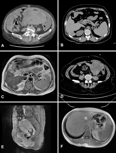
Figure 2 Pathological findings for case 1: (A) (H&E×100), (B) (H&E×200), Microscopically, tumor cells are large and polygonal, with abundant and eosinophilic cytoplasm. The nuclei were lightly stained and appeared round or ovoid, some cells were binucleated, the nucleoli were obvious, mild atypia, and mitotic figures were seen. The nuclei are lightly stained, round or ovoid, with obvious nucleoli, and some of the cells are binucleate. Slight atypia and mitotic figures are visible. The tumor has an infiltrative growth and is poorly demarcated from normal tissue. (C) negative stain for AFP(×100). (D) partial positive stain for Hepatocyte-paraffin 1(×100). Besides, we got immunohistochemical analysis as follows: PAX8(-), P53(-), P16(-), ER(-), PR(-).
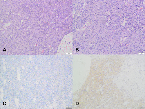
Figure 3 Pathological findings for case 2: (A) (H&E×100), (B) (H&E×200),Microscopically, tumor cells are large and polygonal, with abundant and eosinophilic cytoplasm. The nuclei lightly stained are round, elliptic or irregular in shape, with inconspicuous nucleoli. Some of the nuclei are vacuolated with chromatin squeezed under the thickened nuclear membrane. The nuclei are highly heteromorphic, with active mitoses and some of the cells are binuclear or multinucleate. The tumor has an infiltrative growth and is poorly demarcated from normal tissue. (C) positive stain for AFP(×100). (D) positive stain for Hepatocyte-paraffin 1(×100). (E) negative stain for SALL-4(×100). Besides, we got immunohistochemical analysis as follows: PAX8(-), P53(-), P16(-), ER(-), PR(-).
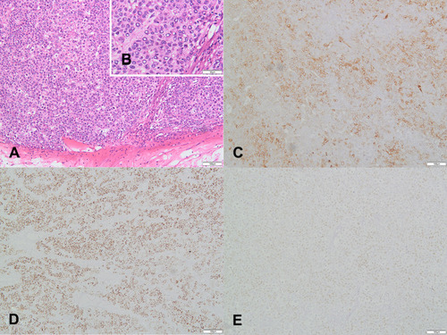
Figure 4 Pathological findings for case 3: (A) (H&E×100), (B) (H&E×200),Microscopically, tumor cells are large and polygonal, with abundant and eosinophilic cytoplasm. The nuclei were round or oval in shape, varied in size, and some cells were binucleate. The nuclear membrane was thickened, the nucleus was pale stained, the nucleolus was obvious, atypia was not obvious, and mitotic figures were seen. The tumor tissue is poorly defined from normal tissue and shows infiltrative growth. (C) positive stain for AFP(×100). (D) negative stain for Hepatocyte-paraffin 1(×100). Besides, we got immunohistochemical analysis as follows: PAX8(-), P53(-), P16(-), ER(-), PR(-).
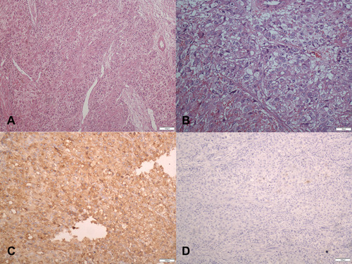
Table 1 Clinical Features, Treatment, and Outcomes of HCO
Table 2 Clinical Features, Treatment, and Outcomes of HCU