Figures & data
Table 1 Primers Used for qRT-PCR Assay
Figure 1 Dysregulation of circ_CORO1C in GC tissues and cells. (A) QRT-PCR assay for the relative expression of circ_CORO1C in GC tissues (n=50) and matched adjacent normal tissues. (B) QRT-PCR assay for the relative expression of circ_CORO1C in GES-1, HGC-27, AGS, MKN45 and MKN74 cells. (C and D) QRT-PCR assay for the relative expression of circ_CORO1C and CORO1C in HGC-27 and MKN45 cells digested with RNase R or not (Mock). *P <0.05.
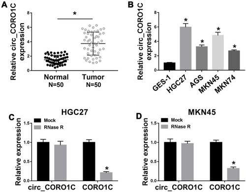
Figure 2 Depletion of circ_CORO1C repressed GC cell proliferation and metastasis, while promoted cell apoptosis. HGC-27 and MKN45 cells were transfected with si-NC or si-circ_CORO1C. (A) QRT-PCR assay for the relative expression of circ_CORO1C in transfected cells. (B) Colony formation assay for the colony formation ability of transfected GC cells. (C and D) MTT assay for the cell viability of transfected cells. (E) Western blot assay for the protein level of Ki67 in transfected cells. (F and G) Transwell assay for the migration and invasion of transfected cells. (H and I) Western blot assay for the protein levels of E-cadherin, N-cadherin and vimentin in transfected cells. (J) Flow cytometry for the apoptotic rate of transfected cells. *P <0.05.
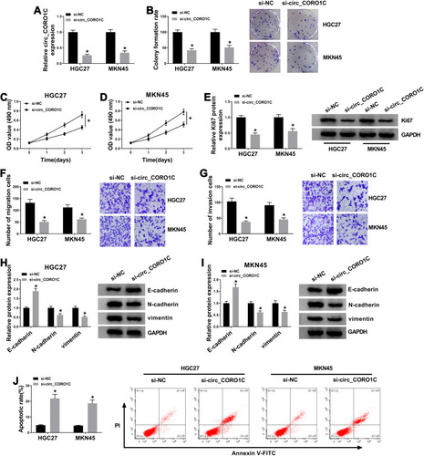
Figure 3 Circ_CORO1C acted as a sponge of miR-138-5p. (A) The binding sites between circ_CORO1C and miR-138-5p, as well as the mutant. (B and C) Dual-luciferase reporter assay for the luciferase intensity of circ_CORO1C-wt and circ_CORO1C-mut in HGC-27 and MKN45 cells transfected with miR-NC or miR-138-5p. (D) QRT-PCR assay for the relative expression of miR-138-5p in HGC-27 and MKN45 cells transfected with si-NC or si-circ_CORO1C. (E) QRT-PCR assay for the relative expression of miR-138-5p in GC tissues (n=50) and matched adjacent normal tissues. (F) Pearson correlation analysis for the correlation between expression of circ_CORO1C and miR-138-5p in GC tissues. (G) QRT-PCR assay for the relative expression of miR-138-5p in GES-1, HGC-27 and MKN45 cells. *P <0.05.
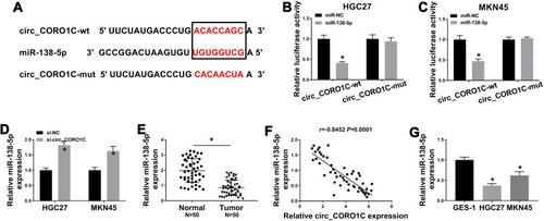
Figure 4 Circ_CORO1C exerted its oncogenic role in GC cells by directly targeting miR-138-5p. HGC-27 and MKN45 cells were transfected with si-NC, si-circ_CORO1C, si-circ_CORO1C+anti-miR-NC or si-circ_CORO1C+anti-miR-138-5p. (A) QRT-PCR assay for the relative expression of miR-138-5p in transfected cells. (B) Colony formation assay for the colony formation ability of transfected GC cells. (C and D) MTT assay for the cell viability of transfected cells. (E) Western blot assay for the protein level of Ki67 in transfected cells. (F and G) Transwell assay for the migration and invasion of transfected cells. (H and I) Western blot assay for the protein levels of E-cadherin, N-cadherin and vimentin in transfected cells. (J) Flow cytometry for the apoptotic rate of transfected cells. *P <0.05.
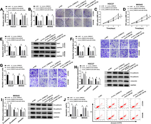
Figure 5 KLF12 was a direct target of miR-138-5p. (A) The binding sites between miR-138-5 and KLF12, as well as the mutant. (B and C) Dual-luciferase reporter assay for the luciferase intensity of KLF12-wt and KLF12-mut in HGC-27 and MKN45 cells transfected with miR-NC or miR-138-5p. (D) QRT-PCR assay for the relative expression of miR-138-5p in HGC-27 and MKN45 cells transfected with miR-NC, miR-138-5p, anti-miR-NC or anti-miR-138-5p. (E and F) QRT-PCR and Western blot assays for the mRNA and protein levels of KLF12 in HGC-27 and MKN45 cells transfected with miR-NC, miR-138-5p, anti-miR-NC or anti-miR-138-5p. (G) QRT-PCR for the mRNA level of KLF12 in GC tissues (n=50) and matched adjacent normal tissues. (H) Western blot assay for the protein level of KLF12 in GC tissues and matched adjacent normal tissues. (I) Pearson correlation analysis for the correlation between expression of KLF12 mRNA and miR-138-5p in GC tissues. (J and K) QRT-PCR and Western blot assays for the mRNA and protein levels of KLF12 in GES-1, HGC-27 and MKN45 cells. *P <0.05.
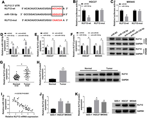
Figure 6 KLF12 could attenuate miR-138-5p-induced GC cell proliferation and metastasis inhibition and cell apoptosis promotion. HGC-27 and MKN45 cells transfected with miR-NC, miR-138-5p, miR-138-5p+pcDNA or miR-138-5p+KLF12. (A and B) QRT-PCR and Western blot assays for the mRNA and protein levels of KLF12 in transfected cells. (C) Colony formation assay for the colony formation ability of transfected GC cells. (D and E) MTT assay for the cell viability of transfected cells. (F) Western blot assay for the protein level of Ki67 in transfected cells. (G and H) Transwell assay for the migration and invasion of transfected cells. (I and J) Western blot assay for the protein levels of E-cadherin, N-cadherin and vimentin in transfected cells. (K) Flow cytometry for the apoptotic rate of transfected cells. *P <0.05.
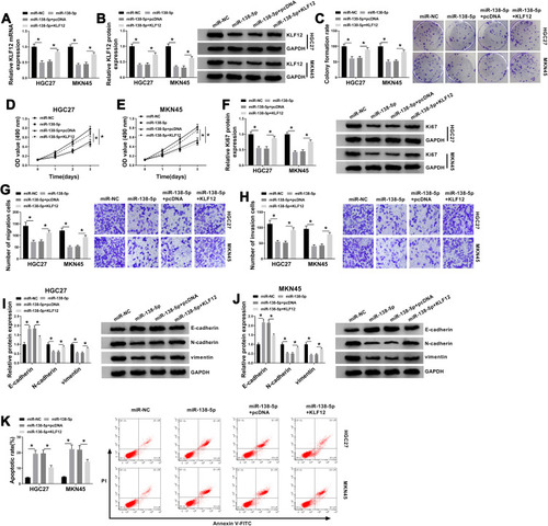
Figure 7 Circ_CORO1C positively regulated KLF12 by sponging miR-138-5p in GC cells. HGC-27 and MKN45 cells transfected with si-NC, si-circ_CORO1C, si-circ_CORO1C+anti-miR-NC or si-circ_CORO1C+anti-miR-138-5p. (A and B) QRT-PCR and Western blot assays for the mRNA and protein levels of KLF12 in transfected cells. *P <0.05.
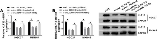
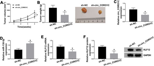
Figure 8 Silencing of circ_CORO1C inhibited tumor growth in vivo. 5-week-old male BALB/C nude mice were subcutaneously injected with MKN45 cells stably expressing sh-circ_CORO1C or sh-NC (n=5). (A) Tumor growth curve of nude mice. (B) Tumor weight in nude mice at 4 weeks post injection. (C and D) QRT-PCR assay for the expression of circ_CORO1C (C) and miR-138-5p (D) in generated tumors. (E and F) QRT-PCR and Western blot assays for the mRNA (E) and protein (F) levels of KLF12 in generated tumors. *P <0.05.