
Figures & data
Figure 1 The examination results of CT, serum tumor marker and FR+-CTC were described in chronological order, included the time before operation, follow-up without medication and after EGFR-TKI treatment. (A and B). CT image of unresected GGO lesions in left upper lobe (LUL) and right upper lobe (RUL) in patients, showing the enlargement and remission of lesions. The yellow arrows in A and B pointed to the location of lesions in the CT images. (C and D). Line chart showed the changes in value of serum tumor marker and FR+-CTC in different periods of time.
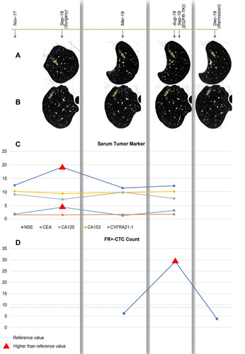
Table 1 Literatures Reporting the Management of Unresected GGO Lesions in SMPLC Patients