Figures & data
Table 1 Basic Characteristics of Meningeal Metastasis Patients
Figure 1 The blood and cerebrospinal fluid of 15 meningeal metastasis patients and 1 brain parenchymal metastasis patient were detected by a panel of 543 cancer-related genes. Heatmap displayed gene mutations in CSF and blood of these patients.
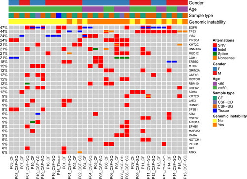
Figure 2 (A–L) A panel analysis of 543 cancer-related genes in blood and CSF in 12 patients with genomic instability. (M–O) A panel analysis of 543 cancer-related genes in blood and CSF in three patients with genomic stability. (P) A panel analysis of 543 cancer-related genes in blood and CSF in a brain parenchymal metastasis patient.
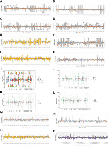
Table 2 Genomic Variation in Cerebrospinal Fluid of the 15 Meningeal Metastasis Patients
Figure 3 (A–F) Copy number variation in blood and CSF in six meningeal metastasis patients with genomic instability. The log2 ratio value is plotted on the y-axis; the x-axis represents chromosomes. Red indicates copy number gain, green indicates copy number loss, and blue indicates no change.
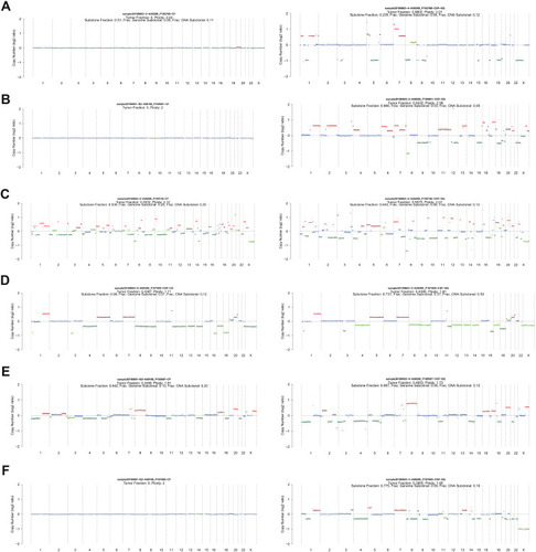
Figure 4 (A and B) Copy number variation in blood and CSF in two meningeal metastasis patients with genomic stability. (C) Copy number variation in blood and CSF in brain parenchymal metastasis case with genomic stability. The log2 ratio value is plotted on the y-axis; the x-axis represents chromosomes. Red indicates copy number gain, green indicates copy number loss, and blue indicates no change.
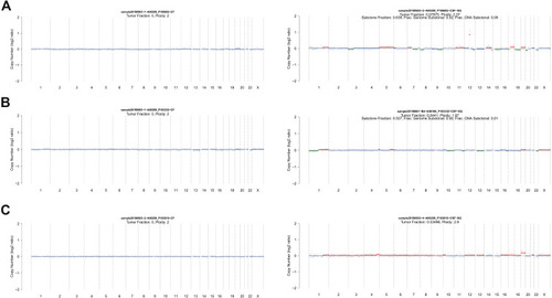
Table 3 Aneuploidy Score of Patients with Genomic Instability or Genomic Stability
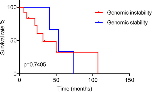
Figure 5 Overall survival analysis between meningeal metastasis patients with genomic instability and genomic stability.