Figures & data
Table 1 Relationship Between XIST Expression and the Clinicopathological Features of WT Patients
Figure 1 XIST lncRNA, YAP mRNA and protein expression in WT blood and tissue samples. (A) ChIP microarray showing differentially expressed lncRNAs in 2 WT and 2 healthy control blood samples. (B) RT-qPCR analysis of 49 pairs of matched WT and adjacent non-tumor samples showing the relative expression levels of XIST. (C) Kaplan-Meier survival curve of WT patients with differential XIST levels. (D) Microarray profiles of differentially expressed mRNA in blood samples of 2 WT patients. (E and F) RT-qPCR analysis of the relative expression of YAP mRNA and miR-194-5p in WT tissues and adjacent non-tumor tissues (n = 49 each for tumor vs normal tissue). (G) Immunohistochemical (400×magnification) and (H) Western blot analyses of YAP protein expression levels in 8 randomly selected pairs of WT tissues and adjacent non-tumor tissues. One-way ANOVA or two-tailed t-test was performed for comparisons between the two groups. **P < 0.01, ***P < 0.001.
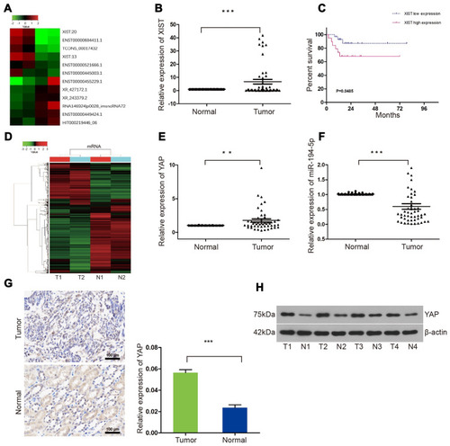
Figure 2 miR-194-5p can bind to both XIST and YAP in WT tissues. XIST lncRNA regulates WT progression through the miR-194-5p/YAP axis. (A) XIST 3ʹ-UTR wild-type (XIST-wt) sequence containing the miR-194-5p binding site and sequence of the mutant (XIST-mut) miR-194-5p binding site. (B and C) Luciferase reporter gene assay (images and histograms) showed lower luciferase activity for miR-194-5p and XIST-wt than XIST-mut (P < 0.05). TRAF6 was used as an internal control to verify the integrity of the luciferase gene reporter assay. (D) YAP 3ʹ-UTR wild-type (YAP-wt) sequence containing the miR-194-5p binding site and sequence of the mutant (YAP-mut) miR-194-5p binding site. (E and F) Luciferase reporter gene assay (images and histograms) showed lower luciferase activity for miR-194-5p and YAP-wt than YAP-mut (P < 0.05). TRAF6 was used as an internal control to verify the integrity of the luciferase gene reporter assay. (G) XIST lncRNA expression and (H) miR-194-5p in WT G401 cells and normal renal epithelial HK2 cells. (I) RT-qPCR analysis of miR-194-5p after transfection of lentiviral XIST, NC, and sh-XIST in WT G401 cells. (J) Western blot analysis showed that YAP protein expression can be regulated by miR-194-5p and XIST. One-way ANOVA or two-tailed t-test was performed for comparisons between the two groups. *P < 0.05, ***P < 0.001.
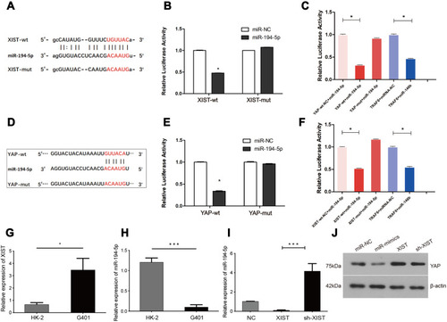
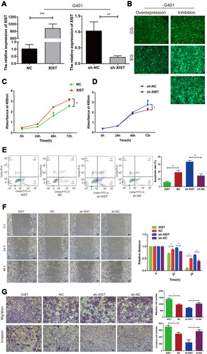
Figure 3 XIST promotes the proliferation, migration, and invasion of G401 cells, and inhibits apoptosis in vitro. (A and B) Stable XIST overexpression (lentiviral XIST and negative control, NC) and XIST knockdown (lentiviral sh-RNA and negative control, sh-NC) were successfully established in WT G401 cells. (C) CCK-8 cell viability assay profiles showed that XIST overexpression enhanced G401 cell proliferation, while (D) XIST knockdown decreased cell proliferation. (E) Flow cytometry showed that XIST overexpression decreased apoptosis while XIST knockdown promoted apoptosis. (F) Scratch assay (100×magnification) (G) and transwell assay showed that XIST overexpression promoted G401 cell migration and invasion in vitro (100×magnification). Three independent replicates were performed. One-way ANOVA or two-tailed t-test was performed for comparisons between the two groups. *P < 0.05, **P < 0.01, ***P < 0.001.