
Figures & data
Figure 1 The first layer of the reconstruction of the posterior wall of total urethral reconstruction of “Sandwich”. (A) The structural location of the medial dorsal raphe (MDR) during the operation. (B) The structural location of the severed end of Denonvillier’s fascia. (C) The severed Denonvillier’s fascia was sutured with MDR.
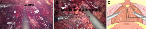
Figure 2 The second layer of the reconstruction of the posterior wall of total urethral reconstruction of “Sandwich”. (A) The structural location of the MDR during the operation. (B) The structural location of the posterior lip of the ladder neck. (C) MDR was sutured with the bladder wall at the back of the posterior lip of the bladder neck.
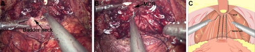
Figure 3 Anastomosis of bladder and urethra of total urethral reconstruction of the “Sandwich”. (A) The structural location of the bladder neck during the operation. (B) The structural location of the urethra. (C) A running urethrovesical anastomosis was performed with the posterior wall suturing from 4 o’clock to 8 o’clock. (D) A urethrovesical anastomosis was performed with the front wall suturing from 3 o’clock to 9 o’clock.
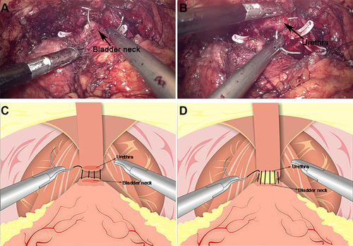
Figure 4 The reconstruction of the anterior of total urethral reconstruction of the “Sandwich”. (A) The structural location of the detrusor apron (DA) during the operation. (B) The structural location of the pubicoprostatic ligament. (C) The pubicoprostatic ligament is sutured with the bladder wall behind the anterior lip of the bladder neck (equivalent to the position of DA).
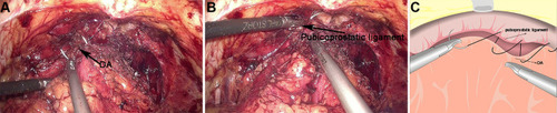
Table 1 Baseline Characteristics of the Patients
Table 2 Operative Date of the Patients
Table 3 Postoperative Data of the Patients
Table 4 Postoperative Urinary Continence Rate of Both Groups