Figures & data
Table 1 Univariate Analysis of Clinical, Vascular and Technical Characteristics of Patients with and without PICC-Related VTE
Table 2 Univariate Analysis of Vascular and Technical Characteristics of Patients with and without PICC-Related VTE
Figure 1 Flowchart showing the final study cohort.
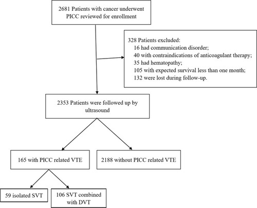
Figure 2 The cumulative incidence of PICC-related VTE. The blue line denotes total incidence of PICC-related VTE; the purple line denotes incidence of isolated SVT; the yellow line denotes incidence of SVT combined with DVT.
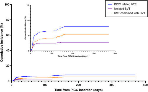
Table 3 Multivariable Logistic Regression of Risk Factors Related to PICC-Related Venous Thromboembolism
Figure 3 Forest plot showing the adjusted odd ratios for risk factors of PICC-related VTE. PICC, peripherally inserted central catheter; VTE, venous thromboembolism.
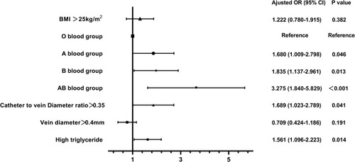
Table 4 Multivariable Analysis of Related Variables in SVT and SVT+DVT Groups
Figure 4 Ultrasound examination of PICC-related VTE. (A–C) show that the basilic vein with PICC-related superficial venous thrombosis was completely occluded and uncompressed without any color signal after 2 weeks of anticoagulation therapy. (D–F) show that the subclavian vein with PICC-related deep venous thrombosis was partially occluded, but was recanalized and filling with blood flow after 2 weeks of anticoagulation therapy.
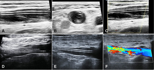
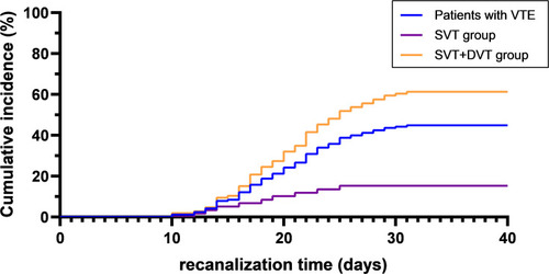
Figure 5 The recanalization time of PICC-related VTE. The blue line denotes all patients with VTE; the purple line denotes the SVT group; the yellow line denotes the SVT+DVT group.